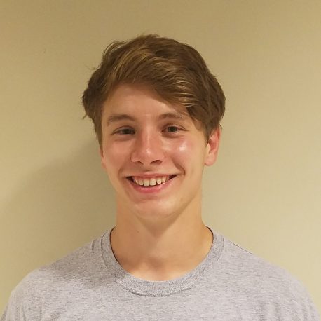
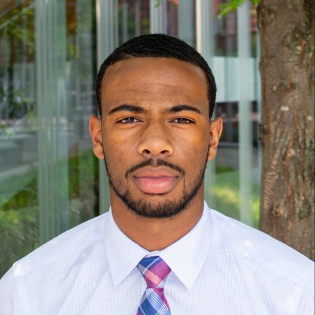
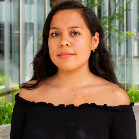
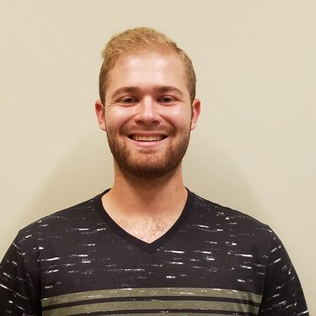
2025 Undergraduates Expanding Boundaries
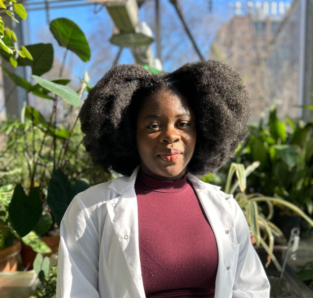
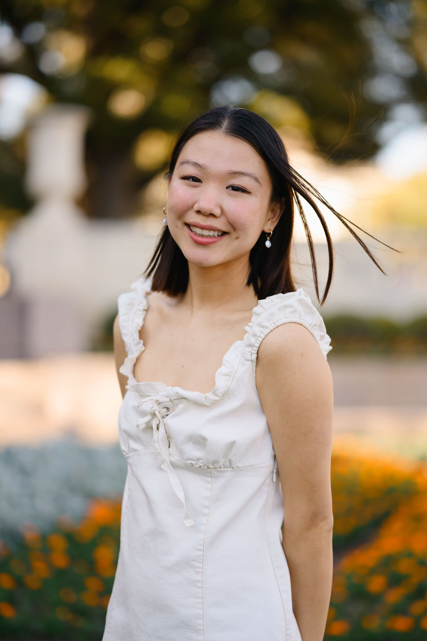
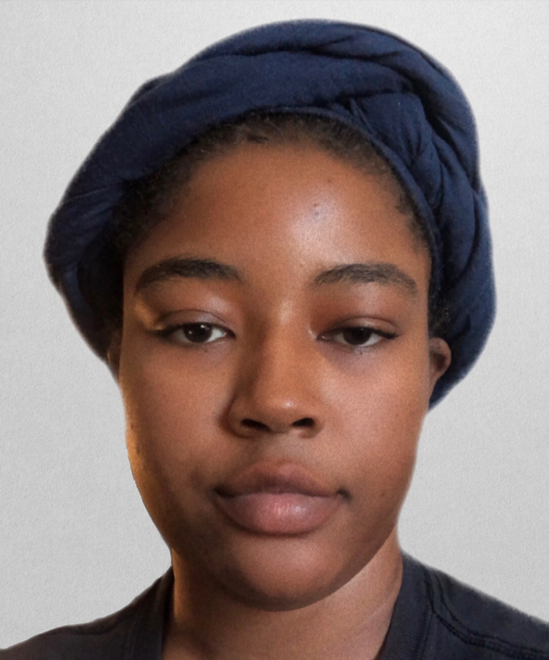
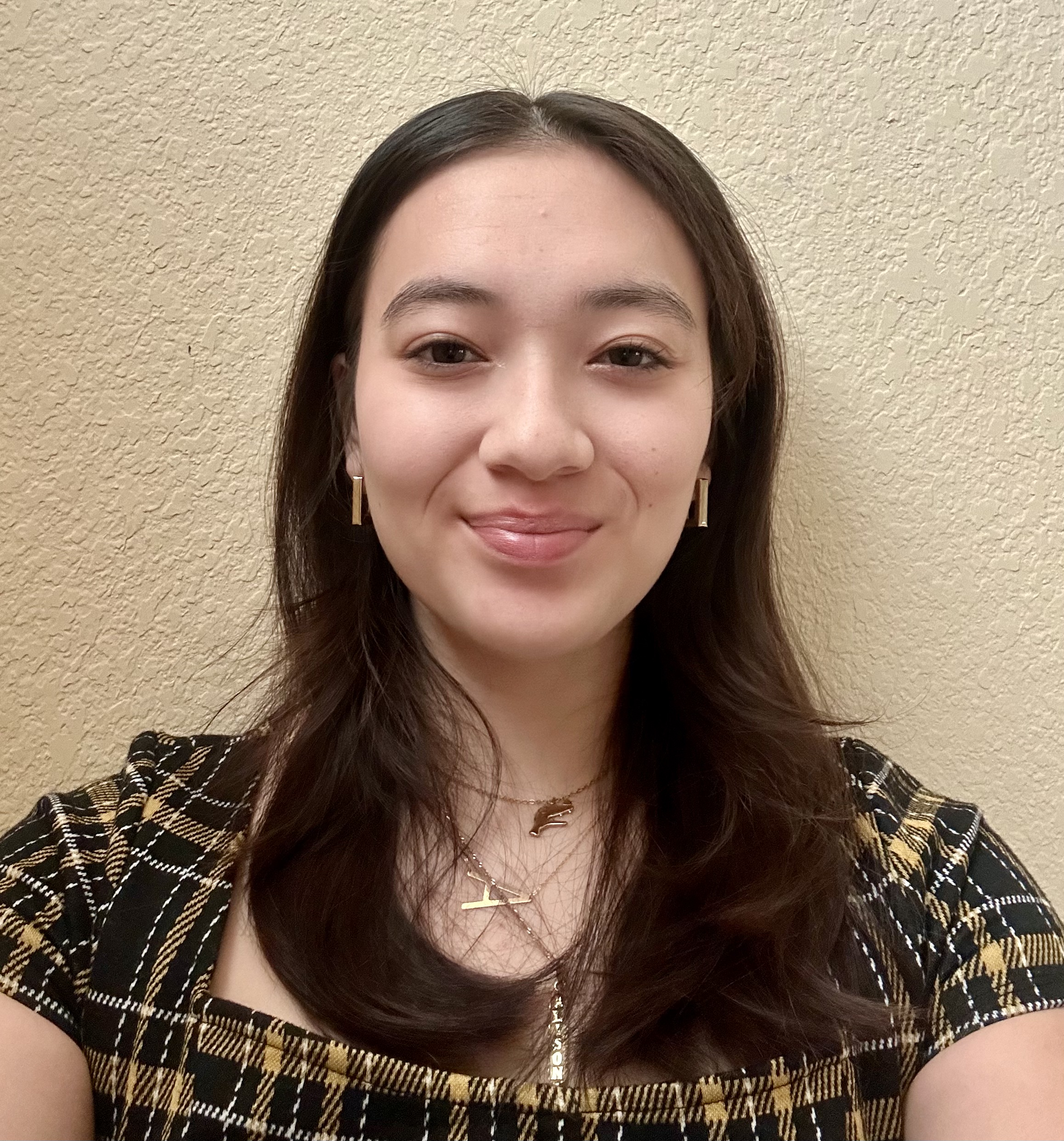
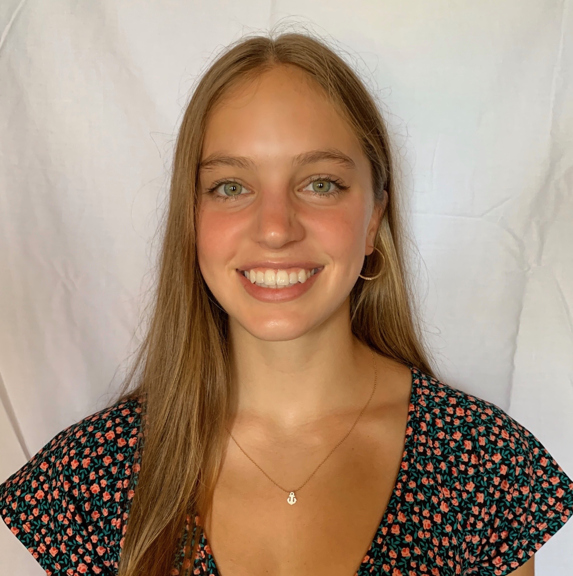
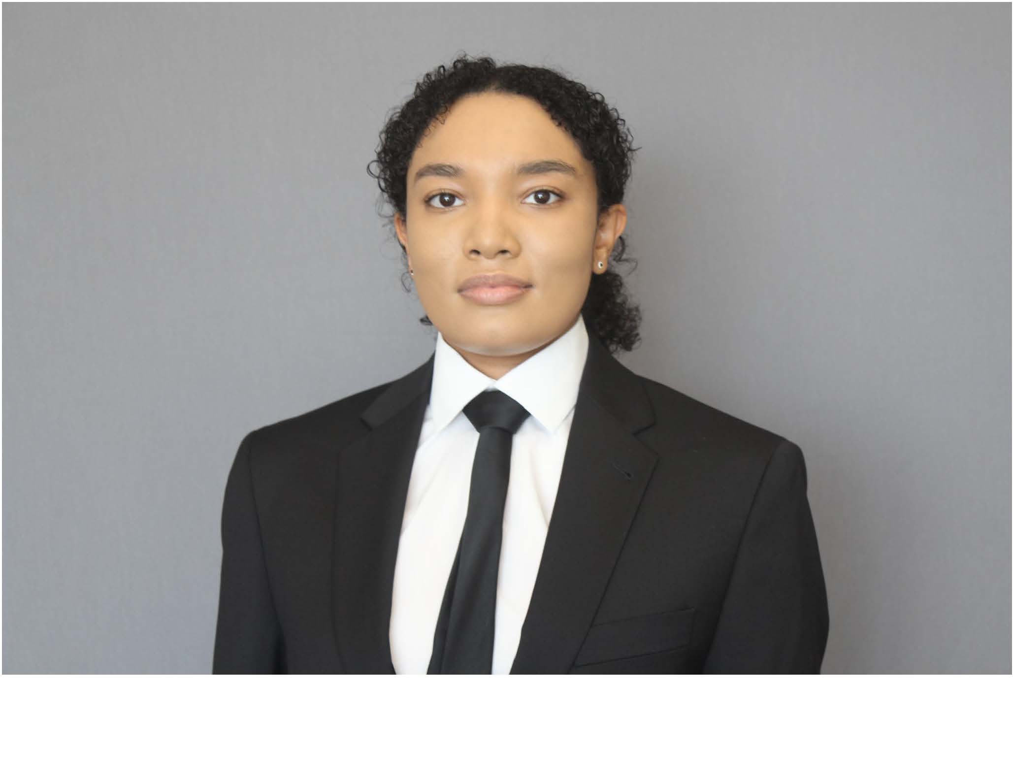
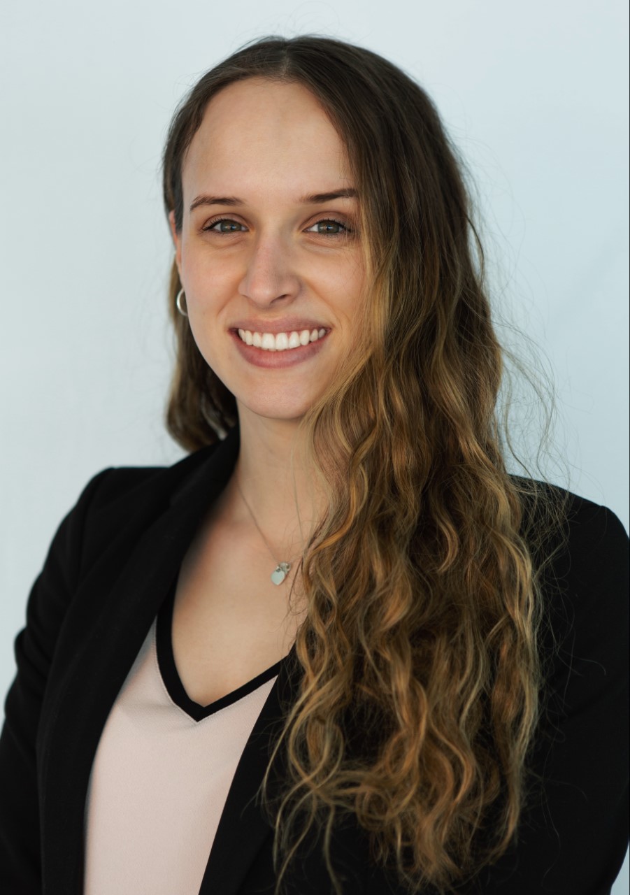
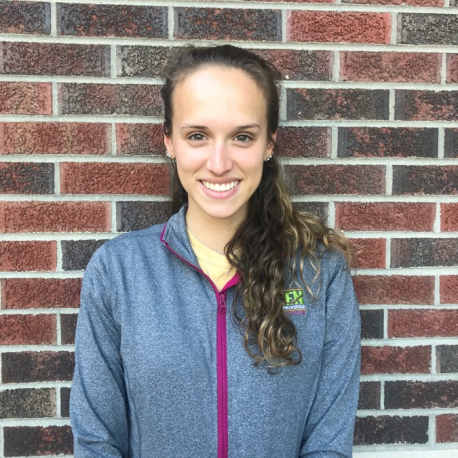
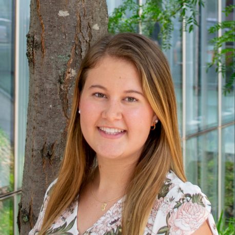
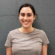
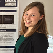
Celine Adomakoh
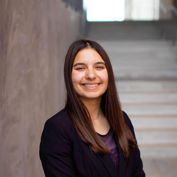
Celine is a Ghanaian-American from Philadelphia, PA. She attends the University of Pennsylvania studying Biophysics pursing a future career in clinical engineering. Her research interests lie at the intersection of bone morphology and biomechanical devices, with a particular focus on musculoskeletal repair and innovation. She is passionate about using medical innovation to improve the quality of life for Black and Brown communities.
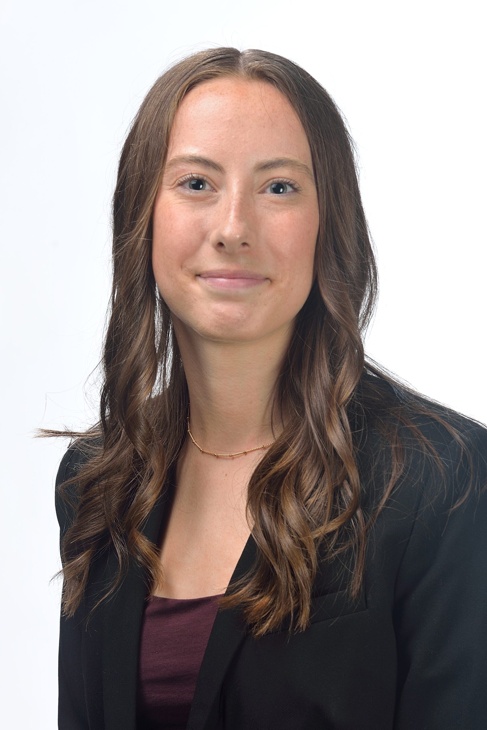
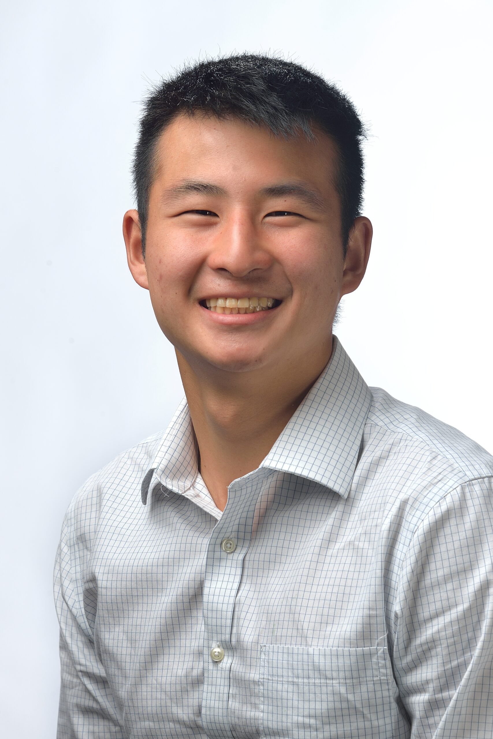
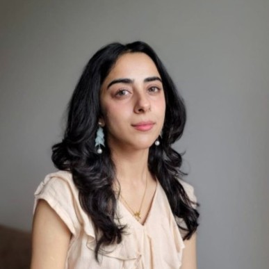
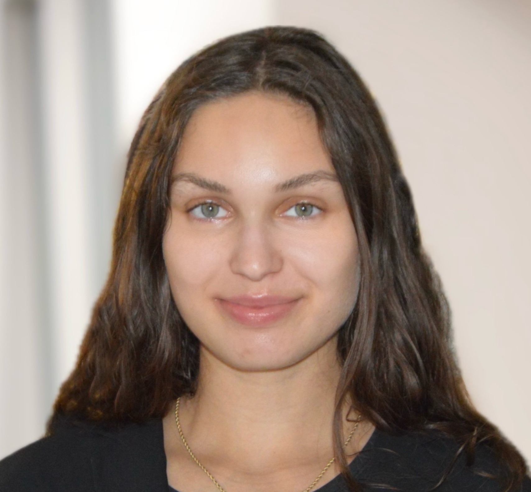
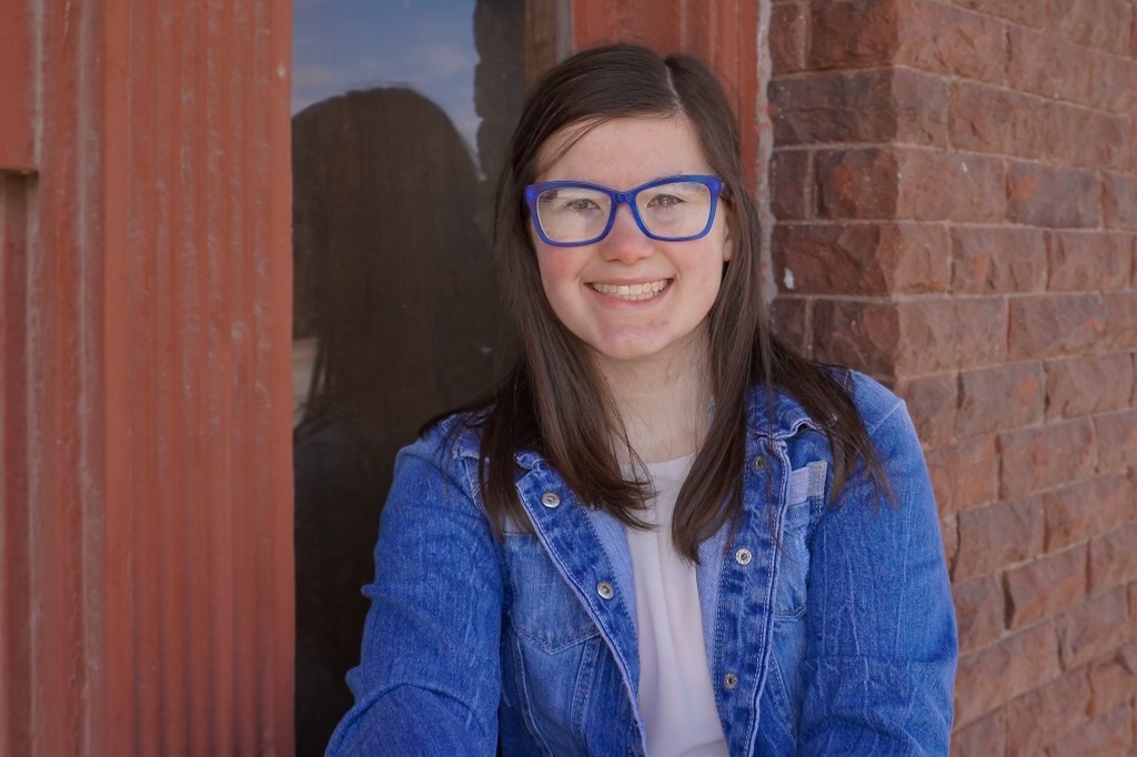
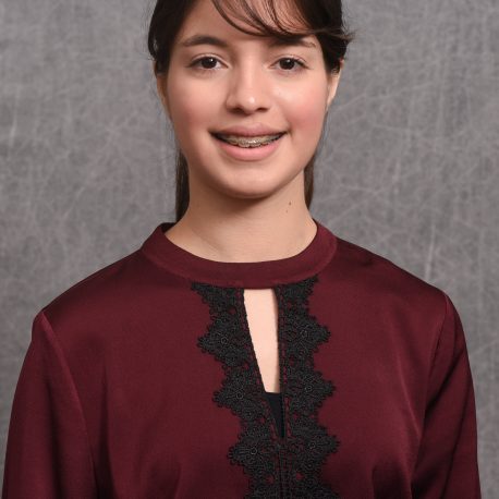
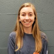
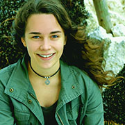
Jasmine Brown
Jasmine is a third-year biomedical engineering student at Georgia Tech. She is interested in regenerative medicine, particularly in cardiovascular tissue engineering. In the future, she hopes to pursue a Ph.D. in biomedical engineering and eventually lead research programs that promote STEM careers among middle and high school students.
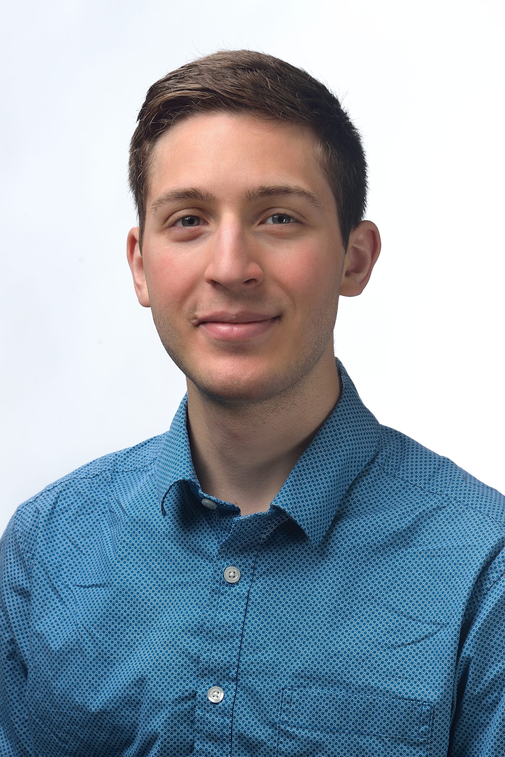
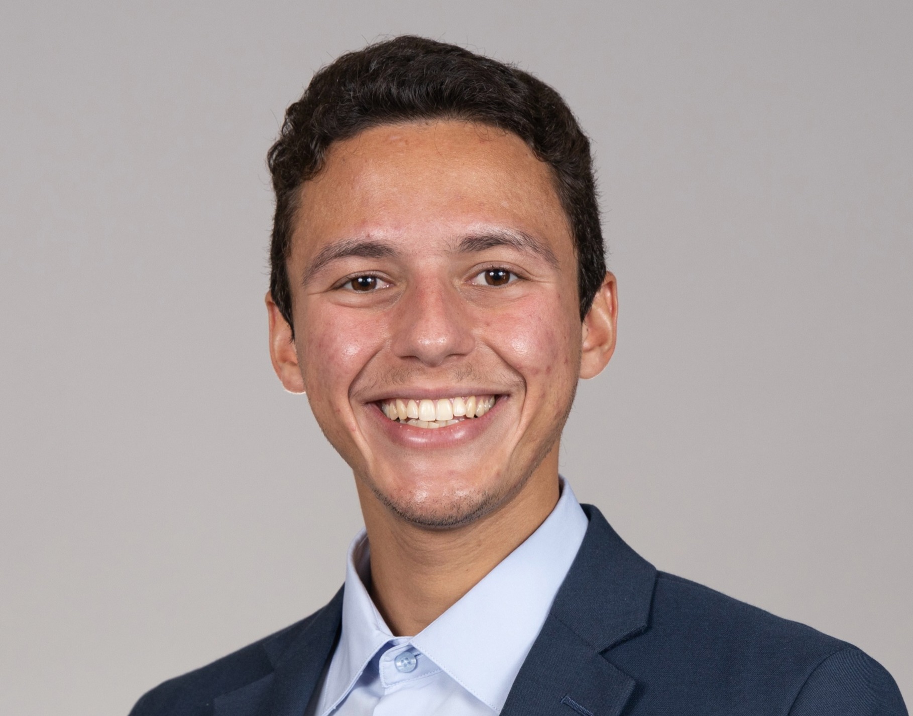
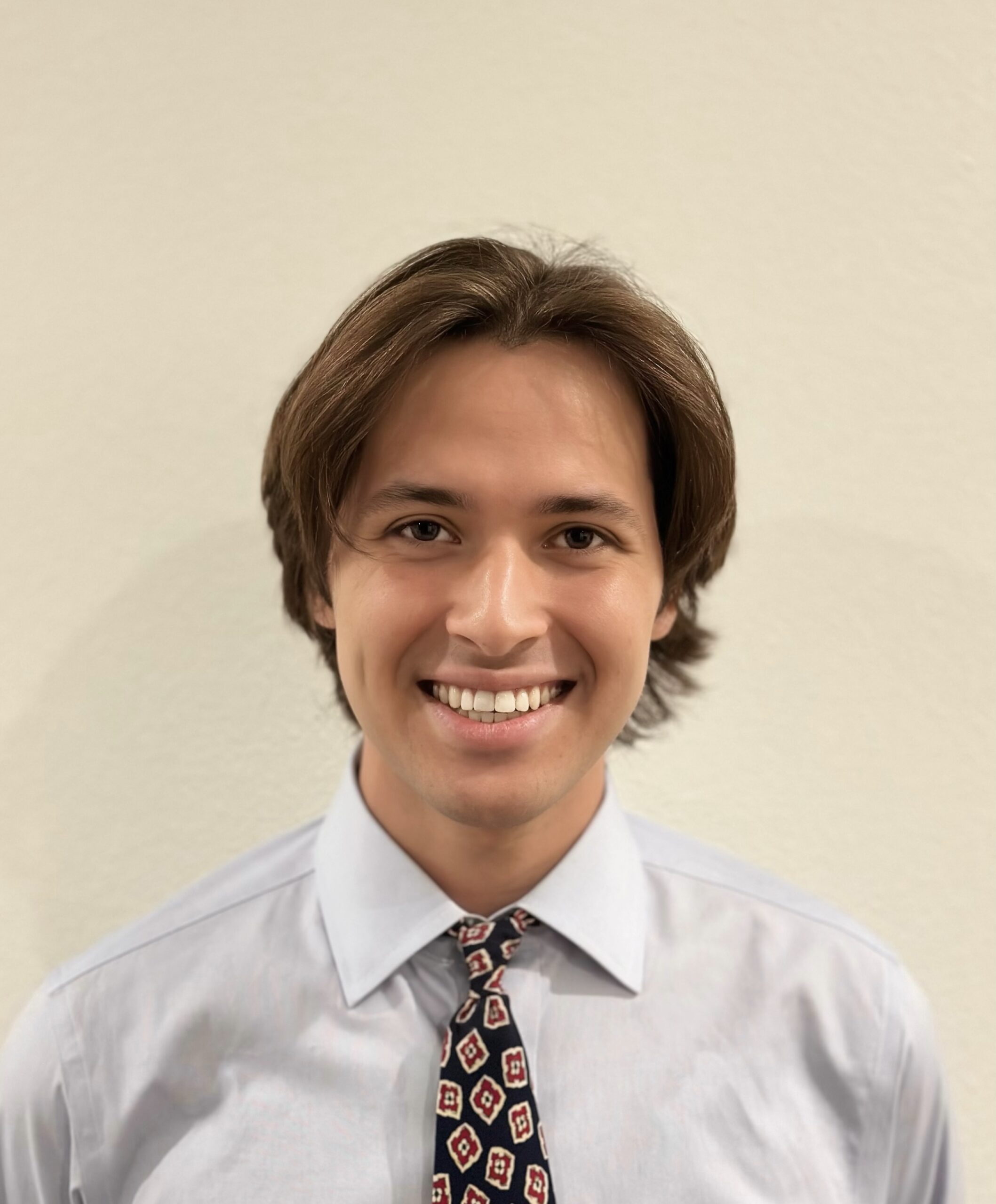
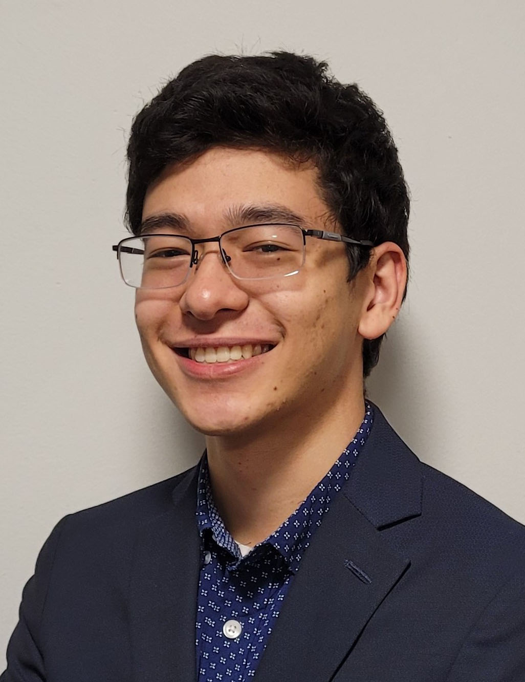
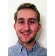
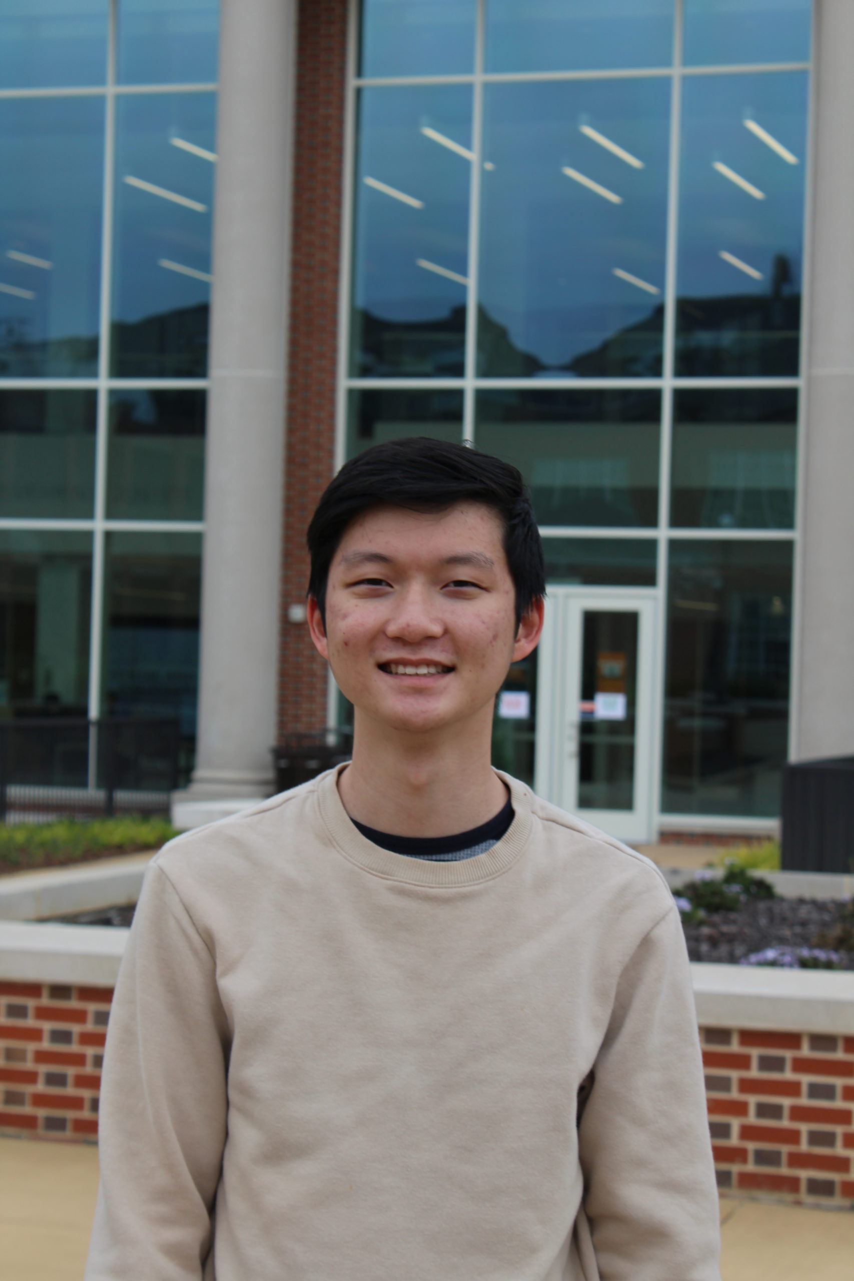
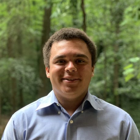
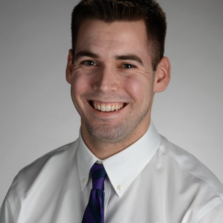
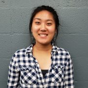
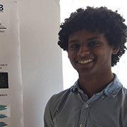
Eric Budu
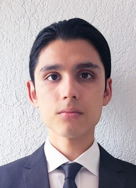
Eric Budu, a Philadelphia native, is a third-year biomedical engineering student at West Chester University of Pennsylvania with interests in biomaterials, orthopedics, nanotechnology, and medical device design. They aspire to obtain a Ph.D. and pursue a career as a Research & Design Engineer in biomedical engineering. In his free time, Eric actively involves himself with the West Chester community through outreach and community service events.
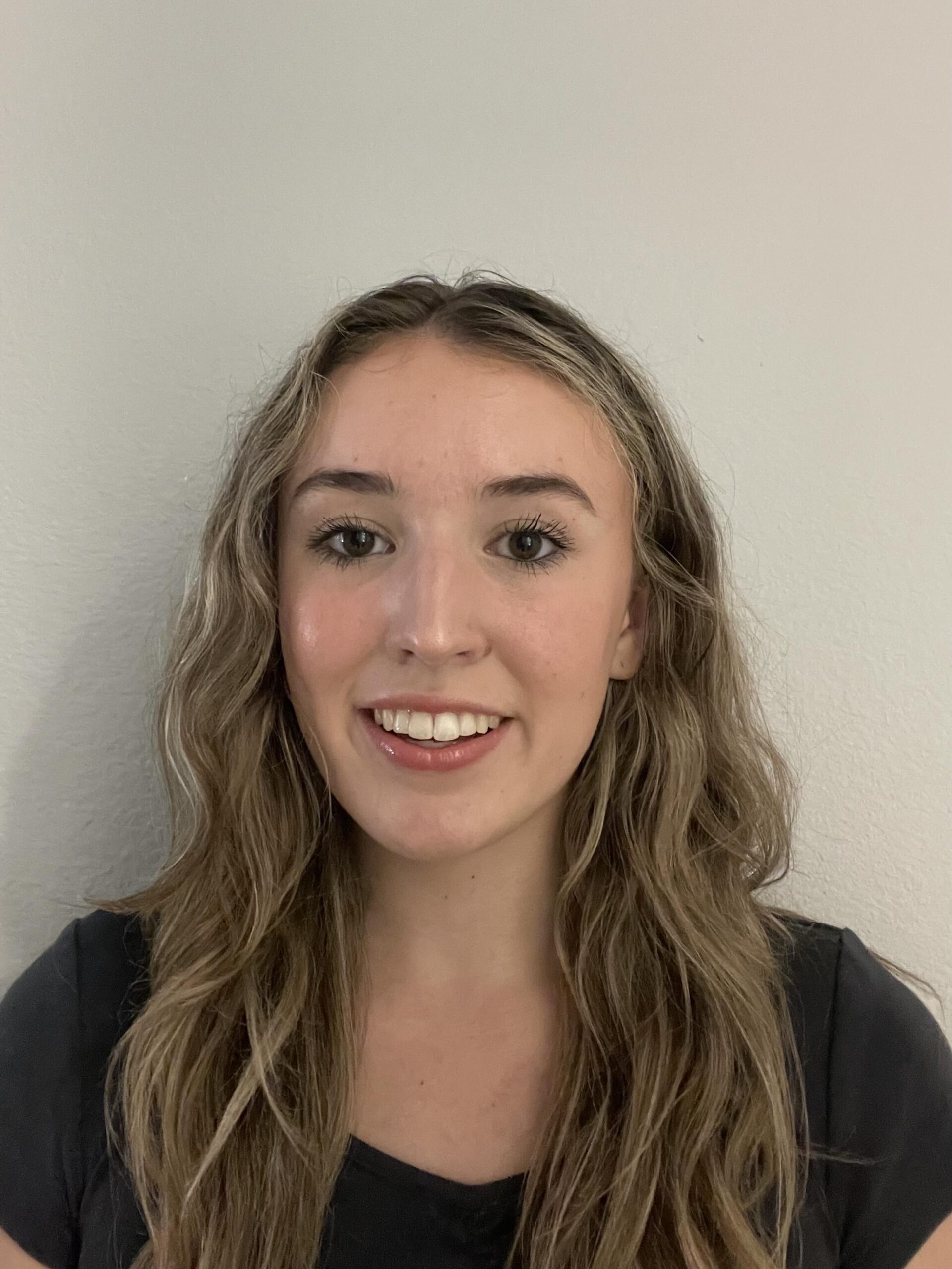
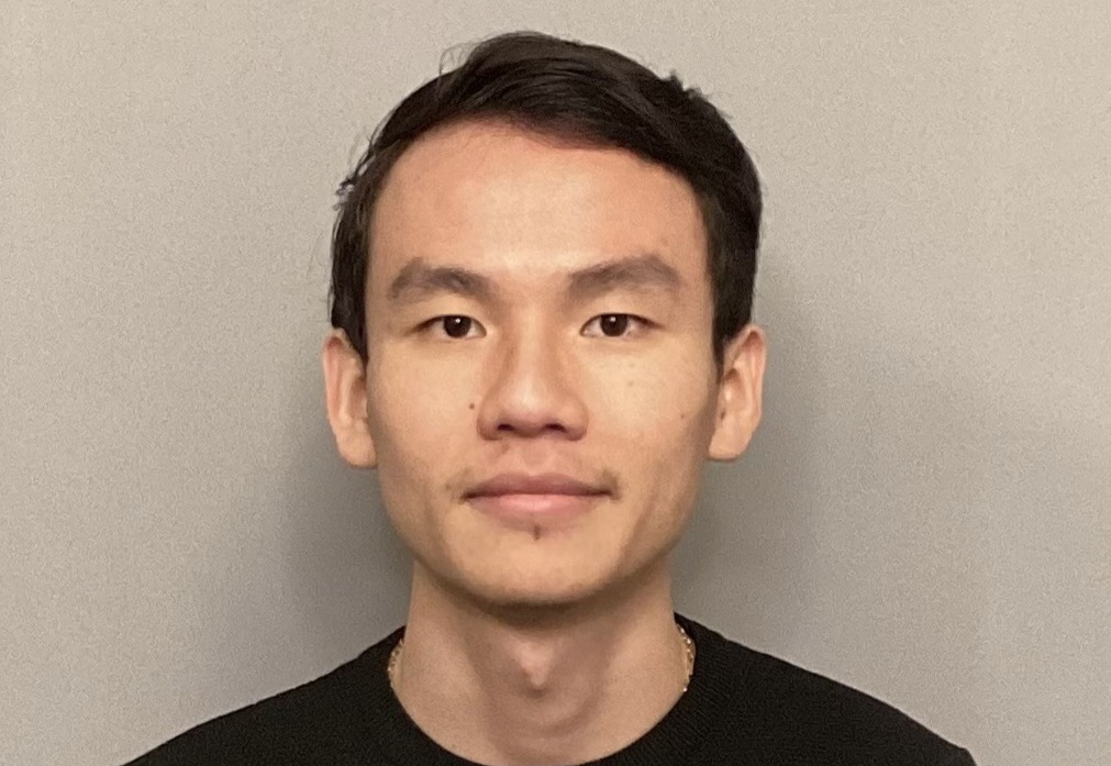
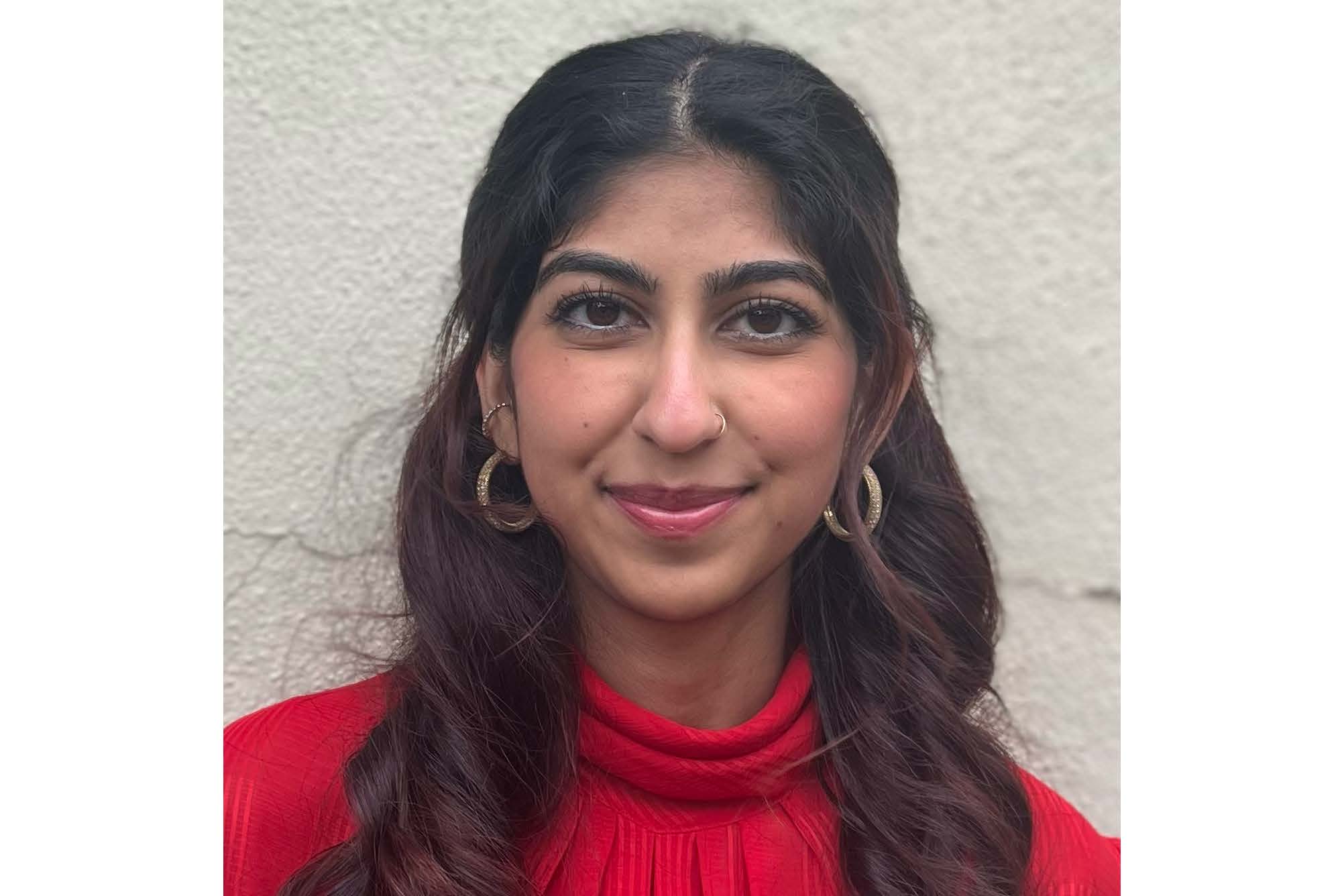
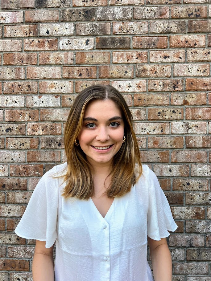
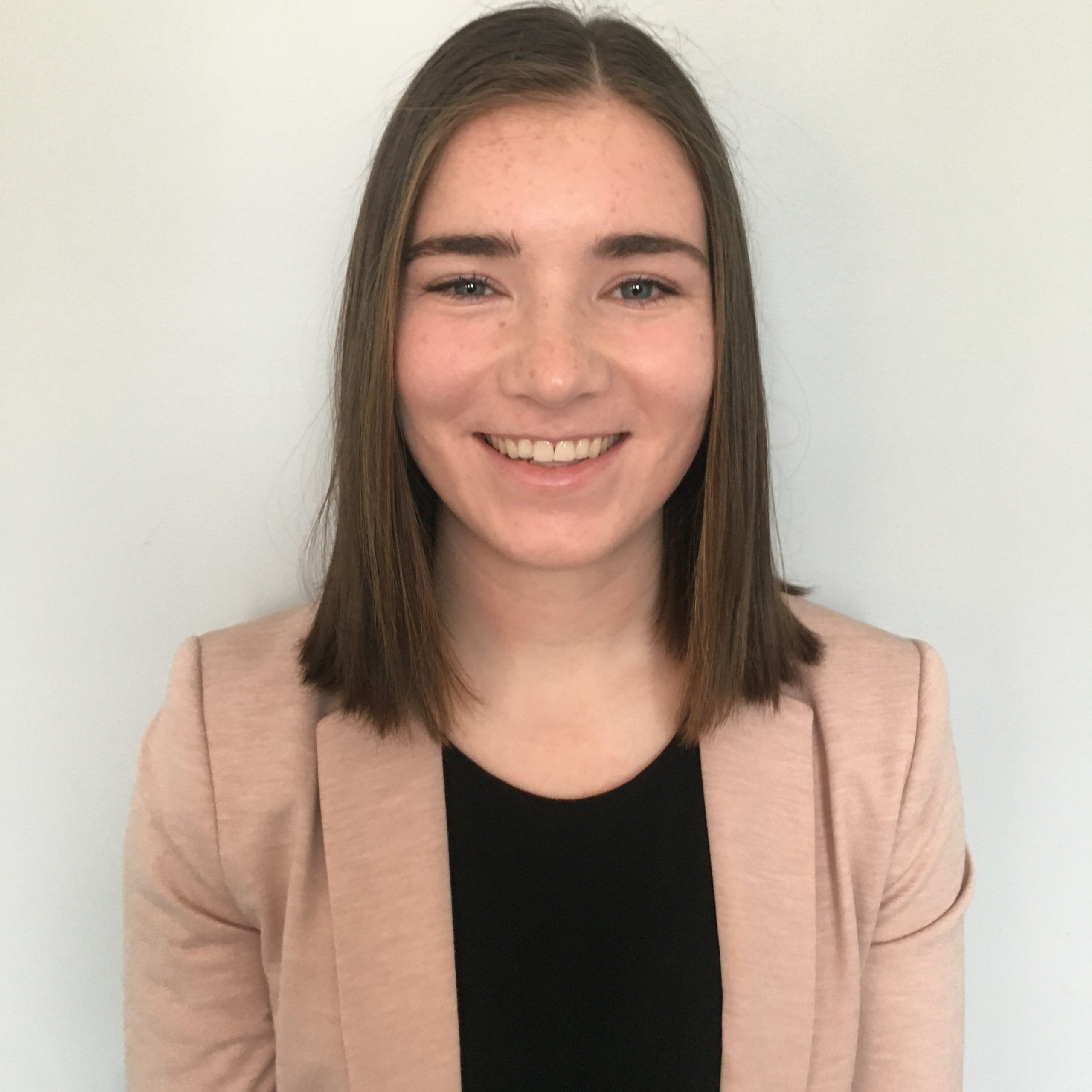
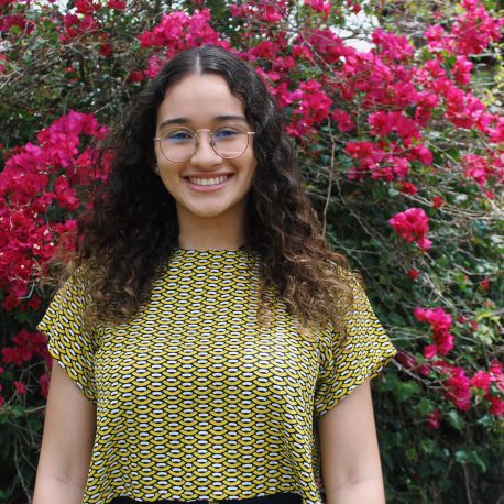
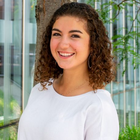
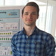
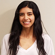
Natalie Calahan
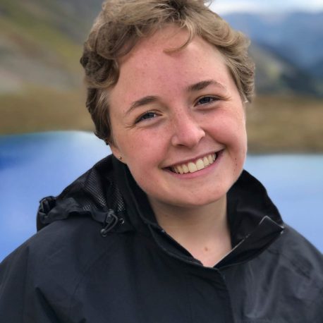
Natalie is a student at Colorado State University studying chemical and biological engineering. She is passionate about tissue engineering and regenerative medicine, with a particular interest in cancer therapeutics. She plans to pursue a PhD in biomedical engineering and aims to contribute to advancements in regenerative medicine and disease treatment.
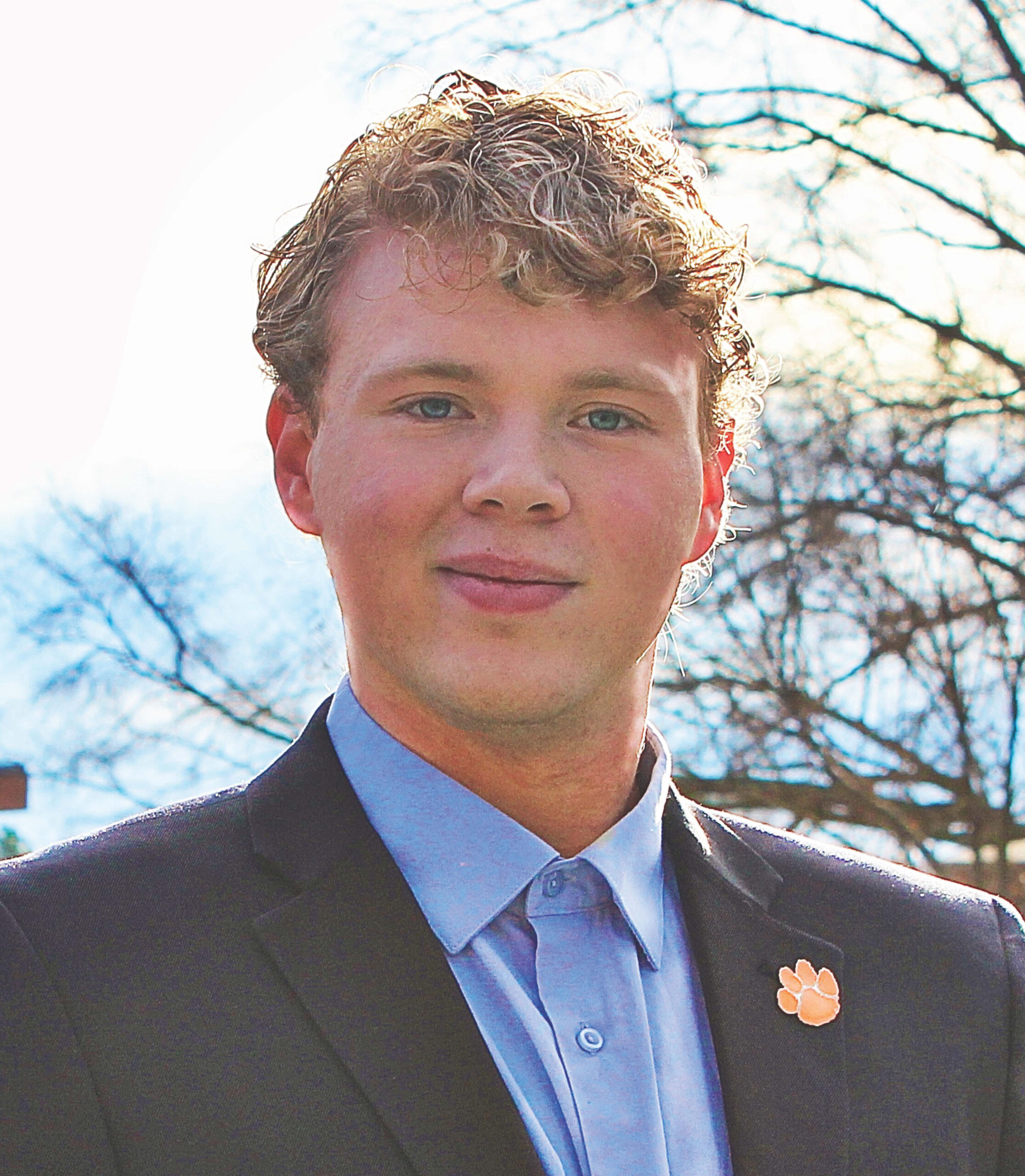
Remy Carlson
Remy Carlson is entering her third year as a biomedical engineering student at the University of Florida. She is actively involved in undergraduate research in a biomechanics lab, exploring innovative solutions in human movement analysis and musculoskeletal diseases. Remy aims to develop technologies that enhance patient care and rehabilitation. Her career aspirations include contributing to cutting-edge research and development in biomedical devices.
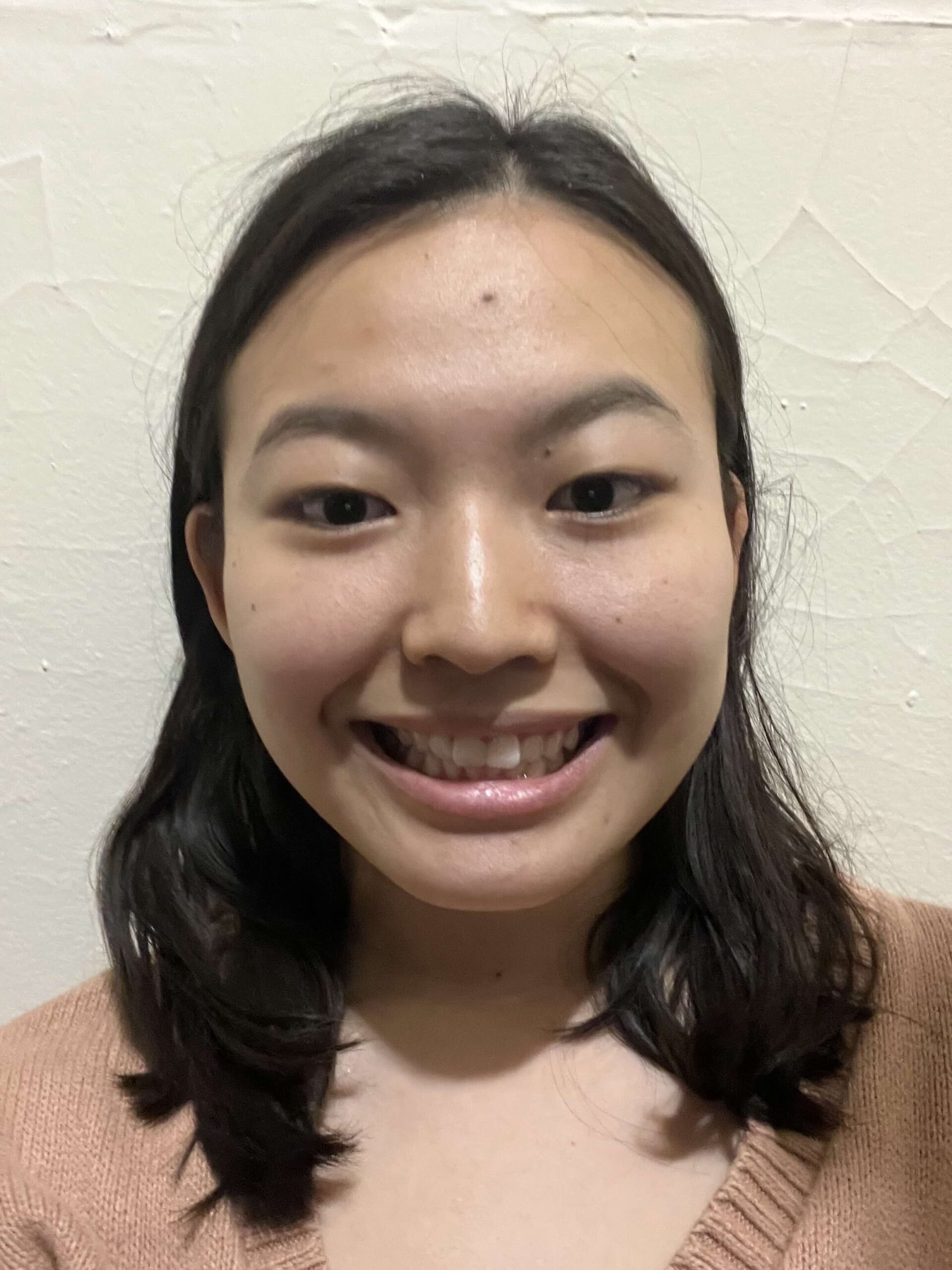
Sowon Kang
Sowon is a rising senior at the University of Texas at Austin majoring in biomedical engineering and minoring in applied statistics. Her research interests include mechanobiology of cancer cells and immunology. After graduation, she wants to go to a graduate school and pursue a PhD in that field.
Tobey Kim
Tobey Kim is a Presidential Fellow and Barry Goldwater Scholar at Bucknell University, where they are a rising junior majoring in Cell Biology and Biochemistry. Their research focuses on the effects of therapeutic ultrasound on chronic wounds. They aspire to pursue an MD/PhD in Biomedical Engineering, with the goal of advancing mechanobiological research and translating discoveries into patient care as a physician-scientist at a medical school.
Angelina Santos
Angelina Santos is a Presidential Fellow and rising junior studying Biomedical Engineering at Bucknell University. She developed a strong interest in mechanobiology during her freshman year while conducting research on the development of a lead-free alternative for low-frequency, low-intensity ultrasound devices designed to stimulate chronic wound healing. Angelina is particularly interested in the intersection of tissue engineering and manufacturing and aspires to continue her research career beyond her undergraduate studies, with the goal of pursuing a PhD.
Madisyn Strange
Madisyn Strange is a second-year Biology major and Presidential Scholar at Hampton University, where she is a member of the Freddye T. Davy Honors College and a player on the HU women’s soccer team. Passionate about biomedical research, she has contributed to studies on hematologic cancers and biomedical technology, with a focus on biomimetic materials for personalized, noninvasive treatments. Her dedication to science and innovation has earned her recognition, including an award for her immunology research at ABRCMS. Aspiring to become a physician-scientist, she aims to bridge engineering and medicine to improve patient care.
Yudith Vazquez-Jacinto
Yudith Vazquez-Jacinto is a senior at Georgia State University majoring in Neuroscience with a minor in Computer Science. She is interested in research at the intersection of Engineering, Neuroscience, and technology, particularly in areas such as computational medicine, neuroengineering, and medical robotics. Yudith plans to pursue a Ph.D. in Biomedical Engineering and aims to contribute to the development of innovative medical technologies that improve brain health and patient outcomes.
Mateo Zevallos
Mateo Zevallos is a rising Senior at The Ohio State University studying Biomedical Science with minors in Anatomy and Portuguese. Mateo has published research in the regenerative biology of zebrafish cardiomyocyte proliferation, aiming to understand the mechanisms behind zebrafish’s ability to regenerate their hearts. Inspired by familial struggles with heart disease, he aims to research mechanisms for inducing heart regeneration in humans to counter the leading cause of death in humans-heart disease. To accomplish that goal Mateo seeks to earn an MD/PhD to become a physician-scientist paving the way for regenerative medicine.
Summer Undergraduates Expanding Boundaries Alumni
2024 Undergraduates Expanding Boundaries
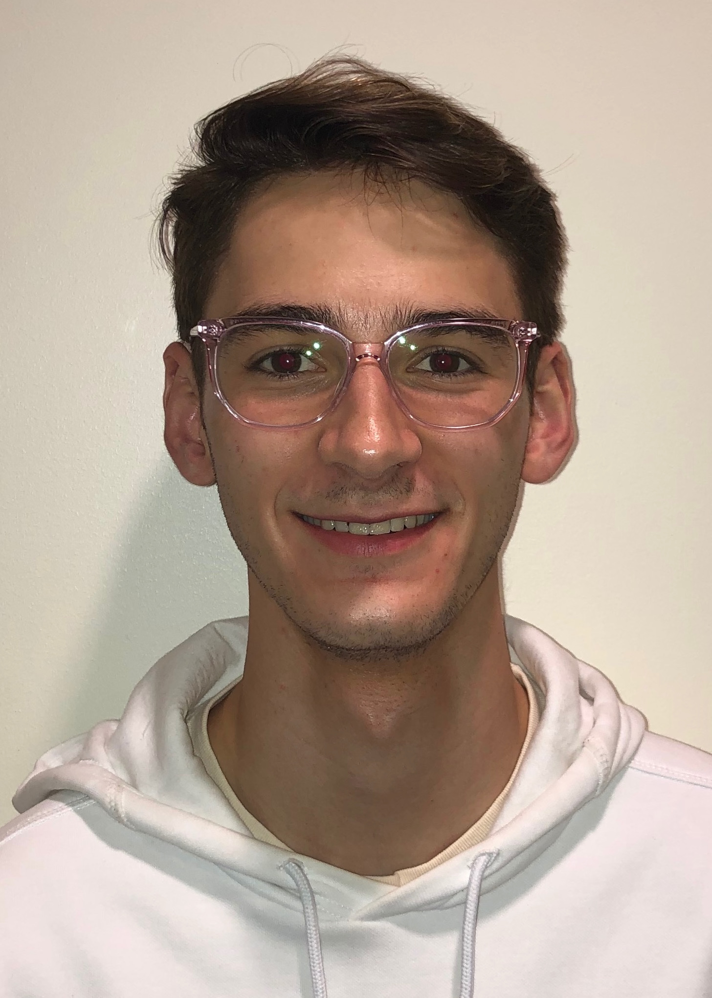
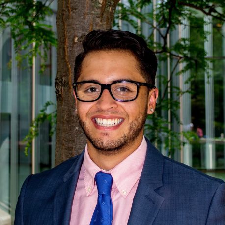
Daniel Aluko
My name is Daniel Aluko and I’m a sophomore student studying Mechanical and Biomedical Engineering at Carnegie Mellon University. My research interests include tissue engineering, orthopedic mechanics, and medical devices, and I hope to pursue a PhD in one of these fields in the future.
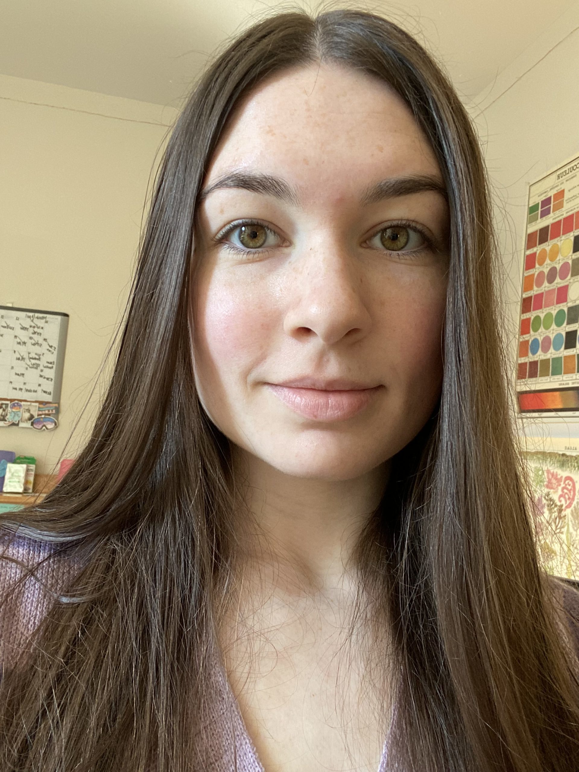
Khalea Avery
I’m a junior biology major hailing from Cleveland, Ohio, currently pursuing my education at Alabama A&M University. My passion lies in the intersection of medicine and research, with a focus on translational science, particularly in the realm of cellular regeneration. Aspiring to become a physician, I aim to contribute to advancements in healthcare through innovative research and clinical practice, with a commitment to serving underserved minority communities.
As of August 2025: In Medical School
Alexandra Goodwin
I am a junior biomedical engineering student from New Jersey Institute of Technology. My research interests lie in genetic engineering, mechanobiology, and the use of CRISPR to study diseases such as cancer. After undergrad, I plan to obtain my PhD in biomedical engineering and hope to continue research while in industry.
Venise Martinez
My name is Venise Martinez. I will graduate from the Community College of Philadelphia with an A.S. in Engineering in 2024. I will transition to Drexel University this Fall as a Junior. I plan on majoring in Chemical Engineering with a minor in Mechanical Engineering. My passion lies in the Biotech industry, and I’m eager to apply my education and skills to contribute to groundbreaking research and innovation.
Winnie Muiru
Hi! My name is Winnie Muiru, I am a rising senior at Bryn Mawr College studying Biochemistry and Molecular Biology. I also conduct research in the Williamson Lab at BMC, which is immunology-focused. I hope to become a physician and to also do research. I love to cook, workout, be in nature and practice hair braiding!
Kehinde Olawepo
Kehinde Olawepo, a rising third-year undergraduate at Alabama A&M University, is pursuing a dual major in Biology and Business, along with a minor in Computer Science, on a Pre-med track. She’s currently participating in the esteemed 2024 CEMB Undergraduates Expanding Boundaries program, where she’s conducting research under the mentorship of Dr. Joel Boerckel in his lab. With aspirations for a combined M.D./MBA degree post-undergrad, Kehinde aims to forge a dynamic path that integrates her passion for medicine and science with a strong interest in business and technology.
Iqra Sajid
I am currently a Junior majoring in Biomedical Engineering at Alabama State University. My professional goal is to aim to contribute to the design, development, and improvement of medical devices, this includes developing robotic systems for surgical procedures. I also hope to seek opportunities to collaborate with physicians and other healthcare professionals to understand clinical needs and challenges and develop solutions that address these needs. My ultimate objective is to initiate the advancement of artificial organ development.
As of August 2025: BME PhD Student at Cornell University
Javier Santiago-Pérez
Hi! I am Javier Santiago-Pérez, a 4th year mechanical engineering student at the University of Puerto Rico (UPR) Mayagüez Campus. Before transferring to Mayagüez, I got a bachelor’s in chemistry at the UPR Río Piedras Campus. My goal is to pursue an MSTP, focused in bioengineering, where I will research biomaterials, tissue engineering and drug delivery. In the long run, I hope to get these exciting developments to my patients, and patients worldwide.
Manav Surti
I am a dedicated junior at the University of Connecticut, majoring in Biomedical Engineering and concentrating in tissue engineering and biomaterials. My academic journey has included substantial research experience in the Hoshino Lab at UConn Storrs, particularly in microscale stiffness testing of biomaterials using mechanical testing tools, lightsheet microscopy, and MATLAB for analysis. As a TRIO Ronald E. McNair Scholar, I am firmly committed to academic excellence and aspire to continue my educational journey by pursuing a master’s or PhD in Biomedical Engineering.
As of August 2025: Biomedical Engineering PhD Student at Cornell University
Maya Taggart Geiss
I’m a junior at Cornell University majoring in biology and food science. In my free time I enjoy biking, writing, and softball. My research interests are centered around bone tissue engineering and cell mechanosensing, and I hope to someday become a physician-scientist specializing in orthopedic surgery!
2023 UExB Alumni
Rachel Callaghan
I'm a Biomedical Engineering major from Michigan Tech, starting my senior year in 2023. My major research experience has been studying types of hydrogels and methods for 3D cell encapsulation and culturing. After graduating I plan on continuing my education in order to earn a PhD and become a college professor. Once I have more control of my research choices I will specialize in studying gynecologic pathology and investigate the under researched disease of endometriosis.
Dongfang (Emily) Chen
Dongfang (Emily) Chen is a rising senior majoring in biomedical engineering at Washington University in St. Louis. She is interested in engineering, biology, and medicine, specifically, the function and treatment of the human body. In the future, she wants to contribute to the development of less invasive diagnostic and treatment methods that can improve quality of life and patient outcomes. She is working in Dr. Jessica Wagenseil’s lab.
Maya Evohr
Maya Evohr is a rising junior majoring in biomedical engineering at Worcester Polytechnic Institute. She is interested in tissue engineering. In the future, she wants to become a professor teaching and running a research lab, and come up with innovative solutions to solve a variety of problems in tissue engineering. She is working in Dr. Nathaniel Huebsch’s lab.
Joshua Gonzalez
My name is Joshua Acosta Gonzalez and I'm a Chemical Engineering undergraduate at the University of Puerto Rico – Mayaguez. I am interested in Biomedical Engineering and aim to obtain a Ph.D degree in this field. My research interests consist of drug delivery, nanotechnology, and regenerative medicine. I aspire to work on creating novel therapies that are accessible to all communities.
Anna Guidry
I am Anna Guidry, a senior at Louisiana State University. I am studying Biological Engineering and exploring how plant based materials can improve wound healing. I am quite interested in the field of tissue engineering. I plan on getting my PhD after graduating in tissue engineering or a similar field.
Lucy Hederick
My name is Lucy Hederick, and I am a junior studying biology and political science on the pre-med track at Northwestern. I am interested in orthopedic research and have spent two summers investigating mechanobiological mechanisms for joint healing. In the future, I hope to attend medical school and eventually become an orthopedic surgeon.
Daichi Kobayashi
Daichi Kobayashi is a rising junior at Washington University in St. Louis. He is pursuing a dual degree in Chemistry and Applied Science (Chemical Engineering). He wants to research the alignment of cellulose nanocrystals (CNCs) in protein nanocomposites for the purpose of developing next-generation nanocomposites to be used in applications from tissue engineering to food packaging. He is working in Dr. Marcus Foston’s lab.
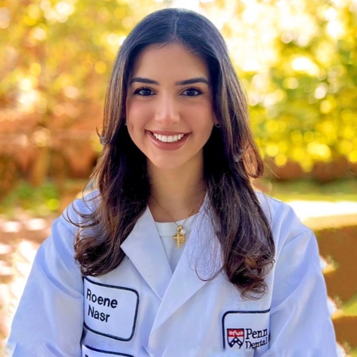
Roene Nasr
Roene Nasr is a third-year undergraduate student at the University of Pennsylvania's College of Arts and Sciences, where she is pursuing a degree in Neuroscience (BS) and minors in Chemistry and French. She is a participant in the 2023 CEMB Undergraduates Expanding Boundaries program, which has allowed her to further her engagement in research on regenerative dentistry, working under the mentorship of Dr. Kyle Vining. In her current project, Roene focuses on the regulation of dental pulp stem cells (DPSCs) in fibrotic hydrogels by furthering her knowledge of how inflammatory white blood cells affect DPSCs in such fibrotic hydrogels with defined biochemical and mechanical cues. Her interests in chemistry and French stem from her recognition of how elaborate the world becomes when we adjust our field of view: by zooming in on the effect of individual atoms and molecules on more extensive systems, and synonymously, zooming out on the power of language and communication. Upon completion of her undergraduate studies, Roene intends to apply to dental school to further her knowledge of dentistry and progress toward a career as both a dentist and researcher.
Eriel Odom
I am currently a junior Chemical Engineering major at Tuskegee University. I'm interested in biochemistry and materials sciences, particularly polymers and plastics. After finishing my undergraduate studies, I intend to pursue a career producing biodegradable and environmentally friendly products.
Simeon Williams
I am Simeon Jerome Williams II! I am currently a sophomore at Morehouse College in Atlanta, GA. I am in the Dual-Degree Engineering Program, majoring in Applied physics and biomedical engineering. Some of my research interests include prosthetics, stem cells, immunoengineering, 3D-printed organs, and nanotechnology. Some of my future goals include going to graduate school and possibly pursuing a law and or business degree. While completing my education, as well as after, I plan to give back to my community whether that's pouring into the next generation of black engineers or helping create technology that allows people to live longer and healthier lives.
2022 UExB Alumni
Rachel Achong
Rachel Achong is a biochemistry student at the University of New Hampshire (Class of 2024) interested in the intersection of biomaterials in dentistry and regenerative medicine. Outside of lab, she enjoys modular origami; this ignited her interest in research upon learning about biomaterials and DNA origami, combining her interests in art, design, and biology.
Seth Ack
Seth is a junior Molecular and Cellular Biology major at the University of Puget Sound (UPS) in Tacoma, Washington. He is also participating in the Dual Degree Engineering program at UPS with the goal of obtaining a masters degree in Biomedical Engineering from Washington University in St. Louis. He hopes to pursue a research career in tissue engineering and regenerative engineering. Outside of lab you’ll find Seth playing with his Australian Shepherd, Nash, rock climbing, or reading fantasy novels!
Lauren Brown
Lauren is a rising senior biomedical engineering student at the University of Massachusetts Amherst. She hopes to perform research investigating cell behavior and disease modeling to advance medical treatments. She is preparing for her upcoming honors thesis investigating the effects of natural toxins on embryonic midbrain and hindbrain development. In her spare time, Lauren enjoys going outside, reading, and practicing yoga.
Alexis Coore
Alex Coore is a student at the University of Alabama set to graduate in 2024. She has done research in the field of Biomedical Engineering and so she is interested in pursuing future research in the same area. Alex hopes to attain at least a Masters in her major before deciding on her final career path.
Kelly Garcia
Kelly is a second year student in The College of Arts and Sciences at University of Pennsylvania studying Neuroscience and minoring in East Asia Studies- China. She has always been very interested in academia and has been in it ever since high school. Now, Kelly looks forward to working with the people at CEMB to learn about the intersection of mathematics, biology, physics, and engineering to explore her passions and pursuits.
Jakub Kowchanowski
Jakub is a bioengineering and physics student at Syracuse University. As a scientist, engineer, and artist, he hopes to combine his many interdisciplinary interests to explore new, creative, paths and technologies that will help the lifeforms of this world and the planet itself. Currently, Jakub’s interests are primarily in biotechnology but he is still exploring the vast potentials and fields of science.
Milan Patel
Milan Patel is a 2nd year honors biomedical engineering and computer science student at NJIT. His academic interests include engineering, biomaterials, physiology, and programming. His experiences surrounding these interests, and in classes and research opportunities, have instilled in him leadership, communication and interpersonal skills, and determination. Milan’s long term goals are to work in the drug design and biomaterials industry, using the research skills he picks up along the way.
Joseph Porter
Joseph Porter is an undergraduate at ETSU anticipating graduation in Spring 2023 with a B.S. in Health Science and a B.S.E. in Engineering. His career goals are in the research and development of neuroengineering technologies, especially in the field of brain-computer interfacing. Hee learned about mechanobiology fairly recently, and although it appears that the majority of studies being performed by the CEMB are targeted at muscle, bone, or epithelial tissue research, he is curious as to how the knowledge and practices of the field may apply to neuroscience.
Lucas Sant'Anna
Lucas Sant’Anna is a rising fourth-year undergraduate at Northwestern University majoring in Biomedical Engineering and Physics. His research interests include synthetic biology and biomaterials, targeted drug delivery, computational biophysics, and drug discovery. His previous publication involves optimizing the performance of the cancer killing molecule TRAIL. After Northwestern, Lucas plans to pursue a Ph.D. in BME.
Ellie Tanner
Ellie Tanner is a sophomore from Columbus, Ohio studying cellular and biomolecular engineering at Purdue University. Currently, Ellie is involved in cancer drug development research at Purdue under Dr. Philip Low, and she looks forward to applying the biochemistry knowledge from this important work to other research in the future. After her undergrad, Ellie plans to pursue a PhD in biomedical engineering or biochemistry to facilitate a career in tissue engineering research. The field of tissue engineering is important to her because she believes that engineered tissues and organs have the potential to change lives for those in need of reconstructive surgeries and transplants, and she wants to help make this a reality for those patients.
2021 UExB Alumni
Marysol Chu
Marysol is a rising senior at The City College of New York, majoring in Biomedical Engineering.
Research Abstract:
Mechanical Basis of Kidney Branching Morphogenesis
Marysol Chu Carty, Michael Foster, Aria Huang, Nathan Petrikas, John Viola, Alex Hughes
Branching morphogenesis in the embryonic kidney leads to the formation of the urinary collecting system. Defective branching can lead to complications such as a lack of one or two kidneys, hypoplasia, or higher risk of adult diseases including hypertension. Branching begins with the invasion of the Wolfian duct into the surrounding mesenchymal tissue leading to the development of a tree-like structure. As this process continues, cap mesenchyme cells epithelialize and mature into nephrons in the “armpit” region of the branching tips. Glial cell derived neurotrophic factor (GDNF), is produced by the cap mesenchyme and promotes ureteric bud (UB) migration by binding and activating Ret, a transmembrane receptor that is present in the UB. The Ret-activated cells at the tips of the UB are more motile and responsible for branching. The trunk cells, which are not Ret- activated are less motile and provide structural support for the ureteric tree; however, the mechanical differences in UB cells due to Ret signaling is not fully understood. We investigated how Ret activation affects the forces exerted by ureteric bud-like cells to understand how branching morphogenesis of the UB sculpts the mechanical environment of the developing kidney. Madin-Darby Canine Kidney (MDCK) cells that were transfected with the Ret gene to express the receptor were used to model the UB. These cells were plated on a 4% T and 2.6% C polyacrylamide gel which was embedded with fluorescent beads. The gel was then functionalized with fibronectin to promote cell adhesion. GFRα-1 and GDNF were used to activate the Ret signaling pathway in one group of cells and contractility was compared to cells in basal media. The cells were imaged using confocal microscopy before and after trypsinization to measure bead displacement. The data that was collected was analyzed and used to generate a vector force map. Based on analysis results from published software, MDCK cells with activated Ret exhibited greater displacement of the beads in the gel and therefore exerted a larger force in the leading and trailing edges of the cell.Our data may contribute new insights into the mechanical effects of GDNF signaling in the developing kidney. In addition to the established roles of GDNF activation in cell motility, it could also mediate intracellular forces and local ECM remodeling as evidenced by higher traction strains measured in our polyacrylamide substrates.
Matthew Egner
Matthew Egner is a junior at the Pennsylvania State University majoring in Biochemistry and Molecular Biology. He plans to pursue a MD-PhD after graduation. During his free time, Matthew enjoys playing backgammon, spikeball and hiking.
Research Abstract:
Cell wall patterning in Arabidopsis thaliana roots is influenced by the mechanical environment in agar and a hydrogel-based transparent soil
Matthew R. Egner, Oskar Siemianowski, Charles T. Anderson
Little is known about the mechanobiology of cell wall patterning in plants. Understanding how roots and cell walls respond to the mechanical environment on the cellular level can provide insights for enhancing plant growth and crop yields. We hypothesized that roots encountering harder media will remodel their cell walls and have different cellulose patterning than roots grown on the surface of or inside softer media. Wild-type Arabidopsis thaliana seedlings were grown on the surface of 0.8% (w/v) agar and inside various concentrations of agar and a hydrogel-based transparent soil composed of alginate/phytagel beads. After 6 days, roots were removed and stained for high-resolution imaging of cell walls with confocal microscopy. ImageJ was used to analyze cellulose orientations. We found that roots grown on the surface of 0.8% (w/v) agar had more deviation of cellulose orientations and a lower anisotropy than those inside 0.8% (w/v) agar. Roots grown inside 0.4% (w/v) agar had less deviation in cellulose orientation and higher anisotropy when compared to those inside 0.8% (w/v) agar. Roots grown in transparent soil composed of 0.8% (w/v) beads contained higher anisotropy than those grown on the surface of agar and in 1.2% (w/v) beads, while those in 0.7% (w/v) beads had the least anisotropy. Together, these results indicate that growing roots inside media might constrain cellulose fibril orientation more than growing roots on an agar surface. When comparing agar concentrations, there was a higher alignment of cellulose in the softer agar than in hard, but in transparent soil, the softest beads had the least aligned fibrils. Future research includes collecting high-resolution in vivo images of cell walls for roots grown inside various mechanical strengths of transparent soil without removing the seedlings for staining.
Bruce Enzmann
Bruce Enzmann is a rising senior at Johns Hopkins University, majoring in Materials Science & Engineering with a concentration in biomaterials. This summer, Bruce is working in Dr. Jason Burdick’s Polymeric Biomaterials Laboratory. After graduation, Bruce plans to pursue a PhD in biomedical engineering to develop translational regenerative medicine.
Research Abstract:
Modeling of Hydrolytically Degradable, Thiol-Ene Crosslinked Hyaluronic Acid Hydrogels
Bruce P. Enzmann III, Jonathan H. Galarraga, Abhishek P. Dhand, Nikolas Di Caprio, Jason A. Burdick
Degradable hydrogels have been developed for a range of biomedical applications, including model culture platforms to investigate cellular perception and response to extracellular microenvironments, the delivery of therapeutic molecules, and the engineering of tissues. Towards their use, further characterization and modeling of degradable hydrogels is needed, particularly for those that undergo hydrolytic degradation. To address this, hyaluronic acid (HA) hydrogels that undergo tunable hydrolysis were fabricated. HA was first reacted with variable concentrations of carbic anhydride to obtain norbornene-modified HA (NorHACA) with hydrolytically labile pendant groups. NorHACA (14, 30, or 40% modification) was then mixed with photoinitiator and di-thiol crosslinker and crosslinked via thiol-ene photocrosslinking. The storage and compressive moduli of NorHACA hydrogels could be readily tuned via changes in polymer concentration, crosslinker concentration, and degree of modification. To characterize bulk degradation, hydrogels were incubated in phosphate buffered saline at 37 °C, the supernatant was collected over time, and degradation profiles were constructed using a 96-well uronic acid assay. Rate constants describing NorHACA hydrolysis were then extrapolated from degradation profiles by assuming pseudo-first order degradation kinetics and performing exponential curve fits. Thereafter, we developed a MATLAB model to predict hydrogel disintegration times and bulk degradation behavior from gel composition and degradation kinetics. NorHACA hydrogels exhibit modular mechanical properties and degradation behavior, demonstrating promise for future applications in tissue engineering and biofabrication. Further development of our model will simulate the degradation of hydrogels composed of multiple polymers, such that hydrogels may be designed with user-defined mechanical properties and disintegration times.
Michael Foster
Michael Foster is a recent Biology graduate of the Community College of Philadelphia, and will be a rising junior at Temple University. Michael is an undergraduate researcher in Dr. Alex Hughes’ lab, where for the summer he is studying how signaling proteins RET and GDNF affect the mechanical environment of the ureteric bud in the kidney, during a process of branching morphogenesis. Michael is focused on pursuing a medical degree, upon graduating from Temple University. Outside of school his interests are traveling, meeting new people, finance, trying new foods, and lifting weights.
Research Abstract
Effects of RET and GDNF to the mechanical environment of the ureteric bud during kidney branching morphogenesis
Michael Foster, Marysol Chu, John Viola, Zheyuan (Aria) Huang, Nathan Petrikas, Alex Hughes
Branching Morphogenesis (BM) contributes to kidney development where the ureteric bud (UB) and metanephric mesenchyme (MM) form by the UB budding into the MM to begin a growing and branching process into a mature ureteric collecting duct. RET and GDNF are two signaling proteins that help direct this process in the kidney. RET is a receptor located on the surface of the UB which has high activity at the tip and has low activity in the trunk of the UB. The high activity of RET in the tip of the UB causes it to be more motile leading it towards the MM which produces GDNF which is a chemoattractant to lead the outgrowth and branching of the UB. Abnormalities to the mechanical environment can lead to defects to the growth and branching of these structures which can result in congenital defects such as renal agenesis, hypoplasia, or congenital obstructive uropathy. These congenital defects could be attributed to the lack of these two signaling proteins causing abnormalities to the mechanical environment. To understand how RET and GDNF affect how the UB cells extracellular mechanical properties impact BM, we used Madin-Darby canine kidney cells (MDCK) an immortalized cell line that are morphologically similar to the renal distal tubular epithelium which in the kidney has high RET expression. The MDCK cells used have been engineered to express RET is known for increasing MDCK cell motility and migration towards a localized source of GDNF ; so the focus is on how RET and GDNF impacts the cells contractility on substrate. Thus, understanding how RET and GDNF affect the contractility of MDCK cells will help give insight on how this interaction affects the mechanical environment and UB growth during BM in the kidney. This can then lead to finding possible methods to clinically treat the defects and abnormalities associated with BM. 2,500 MDCK cells were seeded on a 4%C bis-acrylamide and 2.6%T acrylamide polyacrylamide gel (PA) that is 30 micrometers in height on top of a microscope slide. PA gels were prepared with a 1/10k concentration of fluorescent microbeads. The gel and slide were assembled in an 8-well cell culture chamber. For adherence, gels were coated with a 100 mg/mL concentration of fibronectin, following the application of sulfo-SANPAH to the gel and then were incubated. MDCKs were seeded onto the gel in DMEM media containing GDNF/Gfra-1 and normal DMEM. Images of the cells before and after being detached with trypsin were taken. Fiji plugin iterative PIV was used to measure and analyze the displacement of the beads, followed by FTTC plugin used to obtain a displacement map. We compared RET transformed MDCK cells with MDCK cells that do not express the RET receptor. The MDCK RET cells expressed greater contractility and adhesion when in GDNF/Gfra-1 media. This was shown when the cells were on the PA gel where they displayed high displacement of the fluorescent microbeads. These results suggest that GDNF and RET impact the ability of MDCK cells to contract. Lastly, these findings contribute to the understanding of how RET and GDNF impact the contractility of UB cells inside of the kidney contributing to the mechanical process of branching morphogenesis.
Diana Cruz Garcia
Diana is a rising senior at University of California Merced, majoring in Bioengineering.
Research Abstract:
Role of Actin Stress Fibers and Rho Signaling on Chondrogenic Gene Expression in ATDC5 cells as a Function of Substrate-Stiffness
Diana Cruz Garcia, Ryan Daniels, Robert Mauck
Articular chondrocytes (ACs) are the sole cell type in articular cartilage. These cells produce the matrix that enables a high compressive modulus and a smooth surface for near-frictionless movement. In healthy ACs, actin filaments are separated from the nucleus in a cortical-like organization and express transcription factors Sox9, Collagen 2 (Col 2), and Aggrecan. However, in diseased/dedifferentiated ACs, numerous large actin stress fibers form that engage the nucleus and there is a consequent decrease in chondrocyte-specific gene expression. Increases in canonical Rho signaling leads to the formation of actin stress fibers which results in a decrease of chondrogenic gene expression. Here, we explore the influence of different substrate stiffnesses on ATDC5 cells chondrogenic gene expression when treated with Rho inhibitors/activators and actin stress fiber promoters. ATDC5 cells were seeded on fibronectin-coated polyacrylamide (PA) gels to test the effects of substrate stiffness (glass, 55 kPa, 5 kPa). Small molecules (LPA: Rho agonist, CT04: Rho inhibitor, Jasplakinolide (Jsp): f-actin promoter) were added and RNA was isolated to assay for changes in gene expression using qPCR. We observed that a decrease in substrate stiffness led to a significant increase in chondrogenic gene expression. Our results support a causal link to decreasing substrate stiffness and actin stress fiber formation with increased chondrogenic gene expression. We found that increasing either Rho signaling, or actin stress fiber formation decreased chondrogenic gene expression on soft substrates. These results could suggest that actin stress fiber formation initiates chondrocyte dedifferentiation when chondrocytes are in a diseased or damaged environment.
Zina Helal
Zina is a rising senior at the University of Texas San Antonio, majoring in Biomedical Engineering.
Research Abstract:
Varying Mechanics and Structure of Polyacrylamide Pillars, a Model for the Bile Duct
Zina Helal, David Li, Rebecca G. Wells
Biliary atresia (BA) is a neonatal disease in which bile ducts become fibrotic and can lead to cirrhosis and death within the first years of life if left untreated. Little is understood about BA pathogenesis, but it is likely that ducts become stiffer as fibrosis develops, which may be a key factor affecting cell mechanics and associated disease development.To further understand how stiffened bile duct tissues may affect behavior of the mesenchymal cells surrounding the lumen, we developed an in-vitro polyacrylamide (PA) hydrogel model of the bile duct. We hypothesized that by varying the stiffness and curvature of PA pillars, fabrication can be optimized to model normal and obstructed interlobular and septal bile ducts. An array of polyacrylamide gel micro-pillars was fabricated through an adapted mold-pattern transfer process. Photolithographic techniques were optimized to generate SU-8 photoresist silicon molds containing holes 500 μm in depth with diameters of 75 μm and 300 μm. PA gels with stiffness chosen to mimic those of normal and obstructed bile ducts (1.1 kPa, 3.1 kPa and 14 kPa) are polymerized over the molds using UV photoinitiation. Pillar geometry measured with epi-fluorescent microscopy verified physiologically relevant formation of micro-pillars with height up to 400 μm. Future cell studies seeding gallbladder fibroblast on the pillars will characterize cell phenotype and alignment as a function of pillar stiffness and curvature. This will provide greater insight into how cells sense and respond to stiffened ducts from the smallest parts of the biliary tree and will further our understanding of possible cellular mechanisms involved in BA pathogenesis.
Katherine Kerr
Katherine Kerr is a rising senior at Purdue University, majoring in Biomedical Engineering. She is working in Dr. Paul Janmey’s lab, measuring the shear rheology of soft biomaterials and determining the effects of extracellular proteins on cell morphology. After graduation, Katherine plans on pursuing a PhD in Biomedical Engineering.
Research Abstract:
Update on Viscoelastic Properties of Tofu as a Phantom for Liver Disease Diagnosis
Katherine Kerr, Paul Janmey
Liver disease is the cause of approximately two million deaths globally each year. Many liver diseases, such as liver fibrosis, are characterized by alterations in the mechanical properties of liver tissues. Magnetic resonance elastography (MRE), a method that combines magnetic resonance imaging (MRI) with mechanical stimulation, provides a noninvasive tool to diagnose and monitor liver disease by measuring the mechanical properties of tissues. To ensure accurate evaluations of tissue properties, materials called phantoms are often used to calibrate an MRI device. Traditional phantoms lack complex structures associated with liver tissue. Tofu offers a phantom candidate that has been shown to reflect the materials properties of liver in MRE better than currently available phantoms designed for MRI. In this work, the rheological properties of commercial tofu were studied at different frequencies using a home-made torsion pendulum. The pendulum was compromised of a ruler sandwiched between two aluminum plates, a labjack to which the tofu was fixed, and a rotary motion sensor. A cylindrical tofu sample was sandwiched between the labjack and plates. Weights were added to the ends of the ruler to change the frequency. The plates were rotated by a small angle and then released. The subsequent oscillation angle was measured and used to compute the viscoelastic properties of tofu over a range of 0.5 Hz to 2 Hz. It was found that tofu exhibited frequency dependent properties, being qualitatively similar to liver properties. Our results suggest that tofu could be used as a tissue mimicking phantom to calibrate and validate MRE results.
Noah Kim
Noah Kim is a rising junior at Auburn University, majoring in Mechanical Engineering and Physics. Noah’s favorite subject is physics and is looking to complete a PhD to eventually work in a lab to conduct physics research. He enjoys hiking, skiing, and plays the drums for his church. Noah also likes to watch professional tennis, but is not a very good tennis player himself.
Research Abstract:
A Computational Analysis of Inertial Loading on the Brain
Noah Kim, Aayush Kant, Vivek Shenoy
Computational brain models allow for safe and cost-effective simulation of traumatic brain injury (TBI). However, brain models must include key features to produce accurate results. Brain properties such as anisotropic white matter and ventricles contribute to concentrated areas of shear stress around the ventricular region. Shear stress along neural tracts causes diffuse axonal injury (DAI) in which axons stretch and lose biochemical processes required for regular function, resulting in cell impairment or death. The purpose of this study was to build upon previous TBI experiments and better understand the results of including key brain structures and properties during inertial loading. Data taken from a diffusion tensor imaging (DTI) experiment for a coronal cross-section of the brain was processed using MATLAB to outline the outer boundary of the brain, interface between grey and white matter, and ventricles. Vectors denoting axon tract direction in white matter were filtered to reveal white matter anisotropy. To examine how much the presence of ventricles affected simulated force distribution, multiple models combining or excluding certain factors were created in COMSOL. Material properties from a previous study were used to model the gray matter, white matter, and intraventricular cerebrospinal fluid. Rapid rotational acceleration and deceleration are predicted to contribute greatest towards DAI, so a time-dependent rotational study using a triangular impulse model with max angular velocities ranging from 20 to 200 rad/s was run on each model. Simulation results revealed that high-stress areas were concentrated at the corners of ventricles. Future work will focus on implementation of white-matter and axon tracts into the model.
Kaitlyn McHugh
Kaitlyn is a rising senior at University of Pittsburgh, majoring in chemistry. Kaitlyn will be pursuing a career as a research chemist after graduating, with interests in the fields of nanotechnology, sustainability, and environmental chemistry. In her free time, Kaitlyn enjoys running, ghost hunting, playing video games, and cooking new recipes.
Research Abstract:
Synthesis, Characterization, and Flow Behavior of Biomaterial-based Microfibers
Kaitlyn McHugh, Ravisara (Ning) Wattana, Chinedum Osuji
Microfibers are a unique class of materials because properties like mechanical strength, elasticity, length, and diameter can be controlled during synthesis, which has led to their use in biological processes like drug delivery and tissue engineering. Synthetic polyacrylonitrile (PAN) fibers are often used due to their strong mechanical properties, but they are less biocompatible than natural substances like cellulose nanofibers and alginate. The goal of this study is to investigate the synthesis, characterization, and flow behavior of nanocellulose and alginate-based fibers and compare with PAN fibers. The fibers were prepared by wet spinning precursor solutions into a coagulation bath of nonsolvent. 3% wt PAN was dissolved in dimethylformamide and extruded into water. Various ratios of alginic acid (AA) to cellulose nanofibers (CNF) were prepared by dissolving in water and extruding into 5% wt CaCl2. The morphology of the fibers were investigated under optical microscope and scanning electron microscope. Mechanical properties of the precursors and extruded fibers, including viscosity and tensile strength were analyzed with rheometer. Our results show that fibers in the target range of 17 to 181 μm can be synthesized for most precursor solutions, but mixtures of AA and CNF were more challenging, as the diameter of the CNF-based fibers was more likely to deviate from the size of the extrusion needle. Adding CNF to the AA solution resulted in non-Newtonian behavior, while other precursors exhibit Newtonian behavior. This indicates that many factors determine the characteristics of the final fibers, including flow behavior, extrusion pathway, and extrusion rate.
Sorina Munteanu
Sorina Munteanu is a rising senior at University of California Merced majoring in Bioengineering. Sorina is working in Dr. Guy Genin’s lab this summer, and in the future, Sorina would like to become a physician scientist and conduct translational research on neurodevelopmental diseases.
Research Project: The Role of Actomyosin Contractility in the Dynamics of Microtubule-Based Fibroblast Protrusion
Elaine Nagahara
Elaine is a rising junior at Johns Hopkins University, majoring in Materials Science and Engineering.
Research Abstract:
Understanding the Role of Microenvironmental Curvature on Cholangiocyte Behavior
Elaine A. Nagahara, Kapish Gupta, Jed-Joan S. Edziah, Jiayi Deng, Kathleen J. Stebe, and Rebecca G.Wells
Studies on cell migration and morphology are often done on planar surfaces even though their environment is geometrically complex. In the body, there are various glands that have unique geometries with specific Gaussian curvatures. However, their effects on cell behaviors are not well understood in part due to the difficulty of replicating environmental curvature. Here, using surface tension properties, we developed two curvature models based on particles of different sizes. Collagen poured over the particles formed spherical caps at the top with positive Gaussian curvature and skirts at the bottom with negative Gaussian curvature. Cholangiocytes were then seeded to investigate how such curvature influences cell migration and morphology. We optimized these models to address three fabrication issues: collagen detachment, collagen contraction, and small particle detachment. Preliminary results show that we fabricated models with collagen-substrate contact angle θ = 12.04° and 10.87° and particle-collagen contact angle φ = 21.92° and 24.35° for the larger and smaller structures respectively. In the larger models, cells were seen protruding and migrating at least 250 um (>10x cell diameter) towards the spherical cap. The cells towards the top displayed circularity and aspect ratio 15% lower and 32% higher, respectively, relative to cells towards the bottom. As epithelial cells attach and move, they become less circular and more morphologically fibroblastic, supplementing the idea that the cells towards the top needed to form more adhesions to stabilize their positions on a slope and form protrusions to migrate.
Observing how cells may have migratory preferences for certain curvatures provides insight into controlling cell organization, especially for simulating glands that naturally have Gaussian curvature.
Nathan Rojas Ocampo
Nathan is a rising junior at Columbia University, majoring in Biomedical Engineering.
Research Abstract:
Relating Cell Type Identification to Label-Free Collagen and Myosin Measures in Developing Heart
Nathan Rojas Ocampo, Karanvir Saini, Manasvita Vashisth, Mai Wang, Dennis Discher
Lamin-A is an important protein that has been previously identified as a mechanosensitive protein located under the nuclear membrane that protects cells from DNA damage. It scales with collagen, the most abundant protein in the extracellular matrix (ECM), which also determines tissue stiffness. Low levels of lamin-A paired with constant acto-myosin stress, leads to increased DNA damage due to mislocalization of DNA repair factors. Accumulation of DNA damage in the cell along with telomere shortening may lead to premature aging. Additionally, DNA damage can cause cell cycle arrest and aberrant heart beating in developing hearts. Alterations in the acto-myosin stress within cells of the embryonic chick hearts can be made pharmacologically using blebbistatin, a myosin inhibitor, that ultimately leads to altered beating patterns of the hearts and stress on ECM collagen. Single cell RNA sequencing (scRNA-seq) provides high resolution gene expression of the hearts which enables cell type clustering, comparison of mechanosensitivity, and identification of DNA damage levels to understand which cell types are most mechanosensitive and susceptible to DNA damage due to low levels of lamin-A.
Mistica Lozano Perez
Mistica is a rising junior at University of Texas San Antonio, majoring in Biomedical Engineering.
Research Abstract:
The Effect of Modulating HIF and YAP on Metabolic Activity of Murine Periosteal Cells In Vitro
Mistica Lozano Perez, Madhura Nijsure, Annemarie Lang, Joel D. Boerckel
After bones undergo fractures, multiple pathways activate to aid the bone to regenerate. These pathways involve both mechanical and chemical stimuli. One pathway is known as the HIF pathway and is characterised by activating in response to low oxygen levels in the environment. This pathway occurs because bone fracturing is accompanied with a disturbance in blood vessels surrounding the site of injury, and thus, a loss of oxygen. During this regeneration process, mechanical cues are known to induce two downstream regulators of the HIPPO pathway: Yes-associated protein (YAP) and TAZ. These regulators aid in cell proliferations and are activated by mechanical cues. Both the HIF and HIPPO pathways activate parallel to each other, so we tested whether they interact with one another by first using small molecules to induce or inhibit HIF or YAP in vitro.
Initial results indicated that Deferoxamine (DFO: HIF stabilizer) affected cellular metabolic activity more than other test groups. Following this experiment, we tested the DFO in different concentrations to observe the cellular response to a gradually decreasing concentration of DFO. These results indicated to us that DFO and therefore HIF strongly modulate the metabolic activity of periosteal cells in vitro. Future experiments will focus more on the precise mechanisms that underlie these observations and the possible interactions between this pathway and the HIPPO signaling pathway.
Elizabeth Seidl
Elizabeth Seidl is a rising senior at Kansas State University, majoring in Biological Systems Engineering with a biological focus. This summer, Elizabeth is working in Dr. Dennis Discher’s Laboratory. After graduation, Elizabeth plans to pursue a PhD in biomedical engineering or bioengineering.
Research Abstract:
Actomyosin and Oxidative Stress’s Effect on Heart Beating and Cell Genome Expression
Elizabeth Seidl, Manasvita Vashisth, Karanvir Saini, Mai Wang, Manu Tewari, Dennis Discher
DNA damage within the cell is increases due to mislocalization of the DNA repair factors leading to accumulation of DNA damage, premature aging, cell cycle arrest and aberrant heart beating. Single cell sequencing allows us to look at the different cell types within embryonic chick hearts and compare their genome expression. Understanding acto-myosin stress and DNA damage’s relationship with aberrant beating, cell cycle arrest and the cellular genome can help us better understand what cell types are most susceptible to DNA damage, and eventual possible premature aging. Hearts were treated with four experimental groups: blebbistatin, blebbistatin + washout, DMSO, and H2O2 to induce oxidative and actomyosin stress. Genomic UMAPs were then created using the Seurat package within R, to visualize genomically similar cell clusters, while biomarker genes were used to identify cell types. A wilcox differential gene expression test was run to compare gene mitotic cell cycle genes. Both CENPF and SMC2 were found to be significantly downregulated in the experimental group H2O2 indicating that DNA damage caused by oxidative stress can cause cell cycle arrest. Blebbistatin treatment was found to alter both beating strain and beating frequency of hearts. Using label-free second harmonic generation (SHG) imaging method, collagen and myosin levels measurements were determined within heart tissue samples in order to compare the acto-mysosin stress effects and ECM collagen levels. compared to DMSO. Label free SHG imaging method produced stronger signal in forward direction as compared to backward direction for myosin fibers present within heart tissues.
Mythili Subbanna
Mythili Subbanna is a rising senior from Scarsdale, NY. She studies Mathematics and Biochemistry at Amherst College in Amherst, MA. For her remote research as part of the Anderson Lab this summer, she will model the mechanical interactions and deformation of guard and pavement cells during stomatal opening. In her free time she enjoys going on runs and writing poems. In the future, she hopes to combine her love of math and life sciences in a way that benefits the public good.
Research Abstract:
Predicting the Turgor Pressure-Driven Mechanical Interactions of Guard and Pavement Cells During Stomatal Opening in Plants
Mythili Subbanna, Yintong Chen, Hojae Yi, Eoin McEvoy, Charles T. Anderson
Stomata are tiny pores on the plant surface that regular gas exchange and thus photosynthesis and transpiration. In Arabidopsis thaliana, stomatal complexes comprise two kidney-shaped guard cells and are surrounded by pavement (epidermal) cells. Stomatal opening and closure are determined by ion and water fluxes into and out of guard cells, and their subsequent effects on turgor pressure and cell volume. However, the three-dimensional dynamics of guard and pavement cells during stomatal opening are still unclear, leaving us with an incomplete picture of stomatal mechanics. Previously, we found that during stomatal opening, guard cells increase in XY area while neighboring pavement cells show no significant change in area. Here, we hypothesized that mechanical interactions between guard and pavements cells affects the efficiency of stomatal opening. To understand how guard and pavement cells mechanically interact during stomatal opening, a 3D stomatal complex model was created and analyzed using Finite Element Modeling, a computational method to obtain an approximate solution to governing models for systems with complex geometry under defined constraints. This model encompasses the anisotropic expansion and volume changes of guard cells during stomatal opening. Currently, result generation is in progress. Greater guard to pavement cell turgor pressure ratios resulting in larger increases in stomatal complex width would indicate that efficient stomatal opening depends on the guard cell pushing into the pavement cells. Simulated ablation, or removal, of pavement cells, is expected to allow for further opening of the stomata, supporting the idea that pavement cell presence plays a large mechanical role in proper stomatal opening. We are currently working to place results from our modeling work in the context of underlying ion and water fluxes to understand how cells respond and adapt to their mechanical environment to allow for stomatal function.
2020 UExB Alumni
Joenid Colón-Mateo
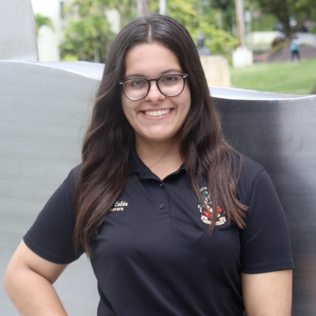
Joenid Colón-Mateo is a rising senior at the University of Puerto Rico at Cayey, majoring in Biology. She is collaborating in Dr. Murat Guvendiren’s lab, which studies the stem cells response to hydrogels with spatiotemporal properties. After graduating, Joenid plans to pursue a PhD in biomedical engineering.
Research Abstract:
Patterning Surfaces for Inducing Cardiomyocyte Alignment
Joenid A. Colón-Mateo, Andrea N. Plaza-Castro, Christian Tessman
Wrinkles are a property found in many biological tissues. Scientists have tried to mimic these surface patterns to understand how cells mechanically interact with their microenvironment. Controlled surface patterns on gels have been shown to affect cell alignment, morphology, gene regulation, and differentiation. Here, wrinkle patterns were fabricated on polydimethylsiloxane (PDMS) substrates to regulate human cardiomyocyte (hCM) alignment, which is important for proper tissue function. PDMS sheets were subject to ultraviolet and ozone (UVO) treatment, with an initial strain of 20%, to form a thin film surface with a higher Young’s modulus than the bulk. Exposure time was modified to determine its effect on wrinkle wavelength, amplitude and film thickness. Analysis of microscope images of the PDMS sheets showed that wrinkle wavelength and amplitude increased linearly with UVO exposure time, and that critical strain decreased linearly with time. The effect of wrinkling on hCM nuclei alignment was also investigated by culturing hCMs on flat and patterned PDMS sheets. Analysis of microscope images of the hCMs showed the average direction of nuclei alignment was similar for both topographical conditions: 84.7±48.0 degrees for flat and 88.1±13.3 degrees for patterned on day 4. However, the standard deviation of nuclei alignment on flat substrates was approximately three times greater than for patterned substrates. This indicates more uniform cellular nuclei alignment on patterned substrates. Development of materials that can mimic surface topography of tissues promises a greater understanding of the morphological response of cells leading to more diverse biomedical applications.
Kathryn Driscoll
Katie Driscoll is a rising junior at Rowan University studying Biomedical Engineering with minors in Arabic, History, and Chemistry, and concentrations in the Honors College and Global Health. She (clearly) has a lot of interests and isn’t sure what she wants to do in the future but will likely be in school for a very long time. This summer she is very excited to be working on identifying scaling relationships of particular genes from cancer patients in the Discher Lab.
Research Abstract:
Scaling Relationships and Patient Prognosis for COL1A1 and ACTA2 in Human Urinary Tract Cancers
Bulk mRNA-seq data obtained from The Cancer Genome Atlas (TCGA) can be used to elucidate gene expression and gene scaling relationships across cancers in order to identify common genes and groups of genes that predict significant survival changes in a cohort of patients. In this analysis, gene scaling relationships among two fibrosis-associated genes of interest (COL1A1 and ACTA2) and four urinary tract cancers were assessed for survival trends across data sets. Bladder cancer (BLCA), Kidney Chromophobe Cancer (KICH), Kidney Clear Cell Carcinoma (KIRC), and Kidney Papillary Cell Carcinoma (KIRP) were chosen for this analysis because they represent a common system and each cancer expressed gene scaling for each gene of interest. For COL1A1, a gene associated with fibrosis and positive survival trends in other cancers1, KIRC and KIRP showed significantly decreased survival of high expressors of the gene and none of the cohorts showed improved survival. Further, in ACTA2, a gene associated with cancer metastasis and fibrosis, only KIRP and BLCA showed significantly reduced survival with KICH indicating non-significant positive survival with high ACTA2 expression. A significant finding in this analysis is that COL5A1, COL6A3, and COL1A2 not only scaled with COL1A1 in all four cancers, but also predicted significantly poor survival with elevated expression in two of the four urinary cancers. These genes are associated with tumor growth and poor prognosis in both urinary cancers and cancers from other systems2,3,4,5. Furthermore, PLN and CNN1 scaled with ACTA2 across all four cancers and indicated negative survival in two of the four. CNN1 has been identified as a possible oncogene in bladder cancer related to poor survival outcomes and PLN has not been previously explored in the context of cancer prognosis6. Taken together, these genes have potential as targets for gene therapies and as prognostic biomarkers. Since many of these genes indicate fibrosis, emerging ultrasound technology could be used as a non-invasive detection tool for urinary cancers7.
- Vashisth, M., Cho, S., Irianto, J., Xia, Y., Wang M., Hayes B., Jafarpour, F., Wells, R., Liu, A., Discher, D. (2020). Scaling concepts in ‘omics: nuclear lamin-B scales with tumor growth and predicts poor prognosis, whereas fibrosis can be pro-survival [unpublished manuscript]. Physical Science Oncology Center at Penn, University of Pennsylvania.
- Di, Y., Chen, D., Yu, W., & Yan, L. (2019, 1 28). Bladder cancer stage-associated hub genes revealed by WGCNA co-expression network analysis. Hereditas, 156(1), 7.
- Kang, C., Wang, J., Axell-House, D., Soni, P., Chu, M.-L., Chipitsyna, G., . . . Arafat, H. (n.d.). 2013 SSAT PLENARY PRESENTATION Clinical Significance of Serum COL6A3 in Pancreatic Ductal Adenocarcinoma.
- Li, J., Ding, Y., & Li, A. (2016, 11 29). Identification of COL1A1 and COL1A2 as candidate prognostic factors in gastric cancer. World Journal of Surgical Oncology, 14(1), 297.
- Liu, W., Wei, H., Gao, Z., Chen, G., Liu, Y., Gao, X., . . . Xiao, J. (2018, 7 30). COL5A1 may contribute the metastasis of lung adenocarcinoma. Gene, 665, 57-66.
- Liu, Y., Wu, X., Wang, G., Hu, S., Zhang, Y., & Zhao, S. (2019, 1 1). CALD1, CNN1, and TAGLN identified as potential prognostic molecular markers of bladder cancer by bioinformatics analysis. Medicine, 98(2), e13847.
- Correas, J. M., Anglicheau, D., Joly, D., Gennisson, J. L., Tanter, M., & Hélénon, O. (2016). Ultrasound-based imaging methods of the kidney-recent developments. Kidney international, 90(6), 1199–1210. https://doi.org/10.1016/j.kint.2016.06.042
- Phillips, J., Pavlovich, C., Walther, M., Ried, T., & Linehan, W. (2001). The genetic basis of renal epithelial tumors: Advances in research and its impact on prognosis and therapy. Current Opinion in Urology, 11(5), 463-469.
- Takahashi, M., Rhodes, D., Furge, K., Kanayama, H., Kagawa, S., Haab, B., & Teh, B. (2001, 8 14). Gene expression profiling of clear cell renal cell carcinoma: Gene identification and prognostic classification. Proceedings of the National Academy of Sciences of the United States of America, 98(17), 9754-9759.
Bruce Enzmann
Bruce Enzmann is a rising junior at Johns Hopkins University, majoring in Materials Science & Engineering with a concentration in biomaterials. This summer, Bruce is working in Dr. Jason Burdick’s Polymeric Biomaterials Laboratory with Dr. Claudia Loebel to analyze patterns of nascent matrix with respect to properties of three-dimensional hydrogels. After graduation, Bruce plans to pursue a PhD in biomedical engineering to develop translational regenerative medicine.
Research Abstract:
Image Analysis to Examine Spatial Properties of the Pericellular Matrix within 3D Hydrogels
Biomaterials, such as hydrogels, can be engineered with biophysical cues that enable the study of three-dimensional microenvironments that simulate aspects of native extracellular matrix and modulate cellular functions such as differentiation and matrix deposition. Recent data showed that the accumulation of deposited matrix in the pericellular region influences the interactions between cells and their engineered hydrogel environment; however, little is known about the spatial localization and density of newly secreted (nascent) matrix at the cell-hydrogel interface. Using a metabolic labeling technique, we fluorescently labeled nascent proteins deposited by bovine chondrocytes within 7 days upon encapsulation in covalently crosslinked 5 kPa and 20 kPa hyaluronic acid hydrogels. To examine spatial properties of these nascent proteins, we used ImageJ to generate nascent protein intensity profiles and developed new analysis tools to quantify nascent protein area and average intensity. Our results show significant increases in nascent protein area and intensity around chondrocytes embedded within 5 kPa hydrogels compared to 20 kPa hydrogels. These findings suggest that secreted matrix within 5 kPa hydrogels distributes further into the hydrogel, whereas the more densely crosslinked 20 kPa hydrogels restrict nascent matrix distribution. Moreover, lower nascent protein average intensity and area within 20 kPa hydrogels indicate that densely crosslinked hydrogels reduce nascent protein deposition. Ongoing work is analyzing the effect of culture time and local mechanical properties on nascent matrix deposition and distribution. We anticipate that these results have implications on hydrogel design for applications in tissue engineering and regenerative medicine.
Samantha Hall
Samantha Hall (Sam) is a rising senior at Bryn Mawr College majoring in Mathematics. Sam is an Accelerated Masters student enrolled at the University of Pennsylvania pursuing an MSE in Systems Engineering. She is from Downingtown, a small suburban town outside of Philadelphia. This summer, Sam is part of Dr. Ravi Radhakrishnan’s lab, which is developing a multiscale computational model for targeting drug-filled nanoparticles to the inflamed lung regions to combat Acute Respiratory Distress Syndrome (ARDS), which manifests in a majority of COVID-19 patients with severe symptoms.
Research Abstract:
Towards a Multiscale Computational Model of the Human Complement System for Predicting Immune Response in COVID-19 Patients
Virion envelope flexibility and receptor spatial arrangement impacts immune modulation, recruitment, and internalization. Given the pandemic’s topical nature, it is advantageous to investigate the mechanics of virion elicited immune response, and how it manifests in patients under pulmonary duress. The majority of COVID-19 deaths occur in patients who have Acute Respiratory Distress Syndrome (ARDS), an acute, diffuse, inflammatory lung injury caused by a variety of insults, most commonly pneumonia, sepsis, trauma, and COVID-19. ARDS affects 200,000 patients each year in the US, has a 40% mortality rate, and occurs in 25% of hospitalized COVID-19-infected patients, yet there are currently no FDA-approved drugs for ARDS. Inflammatory conditions resulting from ARDS are caused by the interaction between the virus and immune cells, namely neutrophils and macrophages. These signaling interactions are primarily mediated via the complement pathway, a part of the immune system that enhances the ability of antibodies to clear pathogens, playing a role in inflammation, host defense, and signaling adaptive immunity. In this project, using methods of systems biology, we look towards signaling models of the complement system to compartmentally understand this complex system. COPASI is a computer software that creates and solves mathematical models, encoding differential equations of biological processes, and was a part of the methodology behind this research. A previously existing computational model involving the enhancement and suppression mechanisms that regulate complement activity provided a template model in Systems Biology Markup Language (SBML) format, a standard form of systems biology XML codes (Liu et al, 2011). Plots were produced of complement regulation with inhibitors under infection inflammation conditions in terms of mediator protein deposition, and of positive feedback amplification of neutrophil activation. Results focused on three aspects: validation of existing data, exploring the amplification modules of the complement pathway, and characterizing emergent properties such as bistable switches regulating the complement cascade. The broader question to be explored in next steps involves how this modeled mechanism would work on the viral surface. Because the complement cascade occurs on the surface of the virus during neutrophil interaction, we believe that the mechanics of the virus, whether it is crystalline or noncrystalline (COVID-19 is noncrystalline), in conjunction with the spatial arrangement of the cascade proteins determine this behavior. Future work involving spatial and stochastic models will involve looking at mechanical and spatio-temporal criterion in virion interaction.
Katherine Kerr
Katherine Kerr is a rising junior at Purdue University, majoring in Biomedical Engineering. She is working in Dr. Paul Janmey’s lab, measuring the shear rheology of soft biomaterials.
Research Abstract:
Viscoelastic Properties of Tofu as a Phantom for Liver Disease Diagnosis
Liver disease is the cause of approximately two million deaths globally each year. Many liver diseases, such as liver fibrosis, are characterized by alterations in the mechanical properties of liver tissues. Magnetic resonance elastography (MRE) provides a noninvasive tool to diagnose and monitor liver disease by measuring the mechanical properties of tissues. To ensure accurate evaluations of tissue properties, materials called phantoms are often used to calibrate an MRE device. Traditional phantoms, such as polymeric gels, can be expensive, difficult to produce at large scales, and lack complex structures associated with liver tissue. Tofu offers a phantom candidate that is cheaper and easy to manufacture. In this work, the rheological properties of two types of commercial tofu, varying in firmness, were studied at different compression levels, making use of a home-made torsion pendulum. The pendulum was compromised of two aluminum plates, a labjack to which the tofu was fixed, and a rotary motion sensor. A cylindrical tofu sample was sandwiched between the labjack and plates. The plates were rotated by a small angle and then released. The subsequent oscillation angle was measured and used to infer the viscoelastic properties of tofu at various compression levels up to fifty percent. It was found that both types of tofu exhibited compression stiffening and shear softening properties, being qualitatively similar to liver properties. Our results suggest that tofu could be used as a tissue mimicking phantom to calibrate and validate MRE results.
Nadja Maldonado Luna
Nadja is a rising senior at the University of Puerto Rico at Mayaguez, majoring in Mechanical Engineering. She is currently working in Rebecca Well’s Lab on the study of lipid droplets and their effect on the development of liver diseases. After finishing her Bachelor’s Degree, Nadja is determined in pursuing a Ph.D. in Biomedical Engineering and continuing a career in academic or industry research. In her free time, she enjoys singing, dancing, and spending time with friends and family.
Research Abstract:
Quantifying repair factors in lipid loaded hepatocytes through image analysis
Nadja M. Maldonado Luna, Mariah A. Turner
Introduction: Hepatocellular carcinoma (HCC) is the most common type of primary liver cancer, often occurs in people with chronic liver diseases, and the presence of cirrhosis is seen as the most significant risk factor [1]. Cirrhosis is characterized by alterations in the extracellular matrix (ECM), including increased deposition of collagen and alignment of the ECM architecture, which drastically increases tissue stiffness [2]. Although HCC is often associated with cirrhosis, it can also arise in non-cirrhotic livers in the context of non-alcoholic fatty liver disease (NAFLD) [3]. Lipid accumulation in the liver cells, characteristic in NAFLD, fills the cell cytoplasm and compresses the nucleus. Based on this morphology, the Wells Lab has hypothesized that the lipid droplets act as a mechanical stress on the nucleus, functioning similarly to tissue stiffness [4]. Nuclear deformation from external sources of mechanical stress, such as migration through constricted environments or culture on stiff substrates, has been shown to increase the frequency of nuclear rupture, leading to depletion of nuclear repair factors and the accumulation of DNA damage [5]. We suggest that deformation due to lipid droplets may similarly lead to the depletion of important repair factors and increased accumulation of double-stranded DNA breaks, which may increase the risk of HCC development in NAFLD livers. The objective of this study was to quantify the impact of lipid accumulation in liver cells (hepatocytes) on the amount of DNA damage repair factor in the cells.
Materials and Methods: Cell culture: Primary human hepatocytes (PHH) were seeded onto collagen-coated polyacrylamide (PAA) gels with storage modulus values of 500 and 10 kPa that are representative of the stiffness of normal and cirrhotic livers, respectively. Cells were also cultured on glass, which is non-physiologically stiff. Fatty acid treatment: After the seeding period, PHH cells were incubated for 48 h in the presence of 400µM oleic acid and 0.5% bovine serum albumin (BSA) solution in DMEM. Oleate is the second most common fatty acid in the human diet and is easily packaged into lipid droplets. Cell staining, microscopy, and image analysis: To identify the lipids and assess gross nuclei morphology, cells were stained with BODIPY and DAPI, respectively. To look at the amount of repair factor, BSA control and oleate-treated cells were stained for Ku80. Cells were imaged using a confocal microscope. Nuclei morphology (area, circularity, and solidity) was analyzed using semi-automated image segmentation and detection of individual nuclei in ImageJ. Ku80 mean intensity and integrated density of the nucleus was measured. One-way ANOVAs were used to test the statistical significance of lipid accumulation on nuclear deformation and on Ku80 staining intensity.
Results and Discussion: Nuclear area, circularity, and solidity for oleate-loaded cells tended to decrease compared to controls, consistent with previous work [4]. Also, Ku80 repair factor means intensity and integrated density decreased with lipid loading of cells seeded on the stiffest substrates. Ku80 mean intensity was associated with the nuclear area, however no strong association was seen for any of the measured shape parameters.
Conclusions: The results of these experiments were consistent with previous work from the lab, showing increased nuclear deformation and compression, and decreased repair factor intensity in oleate-treated cells. Ku80 mean intensity was also correlated with nuclear area, suggesting that nuclear compression may be a contributor to Ku80 decrease in oleate-treated cells. Furthermore, differences between the groups were highest on the stiffest substrates, indicating that some level of tissue stiffening may be required for lipid droplets to act as a mechanical stress. Future work aims to determine if lipid- droplet accumulation increases DNA damage accumulation, through a number of different potential mechanisms.
References: [1] Masuzaki+2009 J Hep. [2] Asselah+2009 Gut. [3] Kanwal+2018 Gastroenterology. [4] Chin+2020 AJP-Gastrointest Liver Physiol. [5] Ivanovska+2019 Biophysical Journal.
Evan Morris
Evan Morris is a rising junior at Washington University in St. Louis, majoring in Biomedical Engineering. He is currently working in Dr. Nathaniel Huebsch’s lab, studying biomaterials and tissue engineering. Evan will be assisting in understanding how mechanical cues within cell-cell and cell-extracellular matrix contacts affect heart development and tissue fate, thus expanding possibilities for 3D modeling and medical interventions for cardiac tissue modeling and regeneration.
Research Title:
Assessing Nucleus Pulposus Phenotype Expression and Extracellular Matrix (ECM) Interaction via Mimetic Peptides in relation to Intervertebral Disc (IVD) Degeneration
[Abstract redacted]Sorina Munteanu
Sorina Munteanu is a rising Junior at University of California Merced majoring in Bioengineering. Sorina is working in Dr. Guy Genin’s lab analyzing oocytes from Xenopus laevis (African clawed frog) to determine the effects ultrasounds have on mechanosensitive ion channels. In the future, Sorina would like to become a physician scientist and conduct translational research on neurodevelopmental diseases.
Research Abstract:
Strain Mapping to Identify the Mechanisms by which Ultrasound Activates Ion Channels
Ultrasound imaging can be used as a therapeutic tool for neurological disorders through stimulation of certain mechanosensitive ion channels. However, the molecular mechanism through which these ion channels are activated is unknown. We hypothesize that ultrasound can cause a mechanical force that deforms the cells, thus activating ion channels. To test this hypothesis, Xenopus oocyte membranes will be analyzed in vitro by confocal microscopy to further elucidate the deformation caused by ultrasound. To evaluate whether ion channel activation relates to mechanical strain, these experiments require real-time estimations of strain fields over the oocyte membrane. We therefore developed a method by which stacks of confocal images could be computationally and quantitatively observed to reveal mechanical changes. Through strain mapping under two- and three-dimensional conditions, the deformation is quantified through displacement tracking. With three-dimensional renderings of the oocytes under strain, normal vectors reveal the expansion or shrinkage of the membrane in the xy plane. Thus, the strain fields could be estimated for subsequent correlation with the activation of mechanosensitive ion channels. We show that the controlled membrane expands laterally and retracts longitudinally as tension is applied at magnitudes that are reasonable estimates of those expected from loading by ultrasound. Elucidating the mechanisms of ultrasound on mechanosensitive ion channels may provide the field of neuroscience with a powerful tool that is comparable to techniques such as optogenetics. The underlying mechanisms of ultrasound could be applied clinically as a therapeutic modality for diseases such as Parkinson’s, depression, and anxiety.
Lily Murchison
Lily Murchison is a Junior studying Engineering in a 3/2 program at Scripps College in Claremont, California. This summer Lily is a part of Dr. Ram Dixit’s lab and is investigating the causes of twisted growth as a result of the spiral1-3 and kclr1 twisted mutants in Arabidopsis. More specifically Lily will be analyzing how these microtubule defects effect cell morphology, growth rate, and microtubule orientation in the root of the plant.
Research Abstract:
The Role of the Microtubule Cytoskeleton in Helical Growth in Plant Roots
Roots are critical organs for finding needed nutrients for plants and using directional growth to avoid foreign obstacles. Plant roots, which are anatomically symmetrical, grow via turgor pressure-driven cell expansion. Plant cells are encompassed by cortical microtubules that guide the deposition of cell wall materials which reinforces the cell wall along the transverse axis in elongating cells and direct cell growth in the vertical direction. Mutations in proteins that interact with microtubules can cause helical growth. The spr1-3 mutant is a point mutation that tracks the growing plus-end of microtubules and produce a right-handed growth pattern. A skewed orientation of the microtubule array has been implicated in causing twisted plant growth in mutants, however the role of skewed microtubules in promoting twisted root growth is not well understood. Furthermore, it is unclear if twisting also occurs at the cell level. Using Arabidopsis thaliana, a commonly used plant for cytoskeletal research, we compared the spr1-3 mutant to wild-type Col-0 to better understand the role microtubule orientation has within this twisted phenotype and what relationship the microtubules have with the morphology of the cells, ultimately aiming to understand how patterns at the cytoskeletal and cell level contribute to this twisted phenotype. To assess if microtubules are skewed prior to the onset of root twisting, microtubule orientation in Col-0 and spr1-3 roots expressing a GFP-TUB6 marker were measured using the FibrilTool ImageJ tool. We found that microtubules in spr1-3 roots adopt a left-handed skew before twisting occurs, suggesting that skewed microtubule array may be what drives the twisting phenotype to occur at the organ level. To assess twisting at the cell level, morphometry of wall lengths and angles were compared between wild-type Col-0 and spr1-3 roots using ImageJ. From this we found that at the cell level symmetry breaks in lateral wall lengths whilst maintaining its symmetry in cell angles. This study will help further inform us how microtubule-level mutations affect cell-level and organ-level directional growth and can be used for future modeling of root skewing.
Andrea N. Plaza-Castro
Andrea is a rising senior at the University of Puerto Rico at Cayey majoring in Biology. She is originally from Ponce but now considers herself from Cayey as well. This summer she is working in Dr. Guvendiren’s Lab. In the future she wants to conduct biomedical research involving stem cells and regeneration in hopes of discovering effective methods to treat metabolic bone diseases.
Research Abstract:
Patterning Surfaces for Inducing Cardiomyocyte Alignment
Andrea N. Plaza-Castro, Joenid A. Colón-Mateo, Christian Tessman
Wrinkles are a property found in many biological tissues. Scientists have tried to mimic these surface patterns to understand how cells mechanically interact with their microenvironment. Controlled surface patterns on gels have been shown to affect cell alignment, morphology, gene regulation, and differentiation. Here, wrinkle patterns were fabricated on polydimethylsiloxane (PDMS) substrates to regulate human cardiomyocyte (hCM) alignment, which is important for proper tissue function. PDMS sheets were subject to ultraviolet and ozone (UVO) treatment, with an initial strain of 20%, to form a thin film surface with a higher Young’s modulus than the bulk. Exposure time was modified to determine its effect on wrinkle wavelength, amplitude and film thickness. Analysis of microscope images of the PDMS sheets showed that wrinkle wavelength and amplitude increased linearly with UVO exposure time, and that critical strain decreased linearly with time. The effect of wrinkling on hCM nuclei alignment was also investigated by culturing hCMs on flat and patterned PDMS sheets. Analysis of microscope images of the hCMs showed the average direction of nuclei alignment was similar for both topographical conditions: 84.7±48.0 degrees for flat and 88.1±13.3 degrees for patterned on day 4. However, the standard deviation of nuclei alignment on flat substrates was approximately three times greater than for patterned substrates. This indicates more uniform cellular nuclei alignment on patterned substrates. Development of materials that can mimic surface topography of tissues promises a greater understanding of the morphological response of cells leading to more diverse biomedical applications.
David Reynolds
David Reynolds is a rising senior currently studying mechanical engineering at Iowa State University. He is an undergraduate researcher in Dr. Amit Pathak’s lab, utilizing computational models to investigate cell migration both under noninvasive and invasive states. After earning his bachelor’s degree, David plans to pursue a PhD in bioengineering.
Research Abstract:
Modeling fibroblast-led collective invasion of carcinoma cells with differing mechanically mediated factors.
David Reynolds, Sabrian Shafi
Prevention of cancer cell dissemination and secondary tumor formation is a major goal of cancer therapy as a majority of cancer deaths are related to metastasis. Metastasis is initiated by the collective invasion of carcinoma cells into local epithelial tissue. Carcinoma cells are able to migrate away from the initial site by following the mechanically-mediated signalings produced by the stromal fibroblasts. Computational modeling offers a simplistic way of isolating the key factors responsible for cancer cell invasion, which is not a possibility in vivo and in vitro studies. By utilizing CompuCell3D software, our model reveals that force-applied fibroblasts with low adhesion energies between fibroblasts and cancer cells demonstrate characteristics of cancer cell invasion as described in the literature. In the future, protease-mediated pathways can also be integrated into the model to better represent the biological phenomena. Using computational models to reproduce cancer cell migration can identify key modulators of its early metastasis.
Sabrina Shafi
Sabrina is a rising senior at The City College of New York studying biomedical engineering. She is working in Dr. Amit Pathak’s lab, where she is analyzing collective cell migration using computational modeling. Sabrina plans to attend graduate school to pursue research in tissue engineering.
Research Abstract:
Modeling fibroblast-led collective invasion of carcinoma cells with differing mechanically mediated factors.
Sabrian Shafi, David Reynolds
Prevention of cancer cell dissemination and secondary tumor formation is a major goal of cancer therapy as a majority of cancer deaths are related to metastasis. Metastasis is initiated by the collective invasion of carcinoma cells into local epithelial tissue. Carcinoma cells are able to migrate away from the initial site by following the mechanically-mediated signalings produced by the stromal fibroblasts. Computational modeling offers a simplistic way of isolating the key factors responsible for cancer cell invasion, which is not a possibility in vivo and in vitro studies. By utilizing CompuCell3D software, our model reveals that force-applied fibroblasts with low adhesion energies between fibroblasts and cancer cells demonstrate characteristics of cancer cell invasion as described in the literature. In the future, protease-mediated pathways can also be integrated into the model to better represent the biological phenomena. Using computational models to reproduce cancer cell migration can identify key modulators of its early metastasis.
Mythili Subbanna
Mythili Subbanna is a rising junior from Scarsdale, NY. She studies Mathematics – while also taking biology, chemistry, and physics coursework – at Amherst College in Amherst, MA. This summer, in the Anderson/Shenoy Lab, she will measure plant cell volumes and model water/ion flow in and out of cells to understand how these processes are influenced by cell wall mechanics, and how they affect cell pressurization and stomatal dynamics. In the future, she hopes to do applied mathematical modeling research, as well as pursue a PhD in biomedical engineering or materials science engineering.
Research Abstract:
Measuring and Modeling How Turgor Pressure-Driven Mechanical Forces Between Guard and Pavement Cells Underlie Stomatal Opening and Closing
Mythili Subbanna, Emily Walter
Stomata are pores in the plant surface that facilitate photosynthesis and transpiration. In Arabidopsis thaliana, stomatal complexes comprise two kidney-shaped guard cells, and are flanked by neighboring pavement cells. The pathways by which cytosolic pressure changes, brought about by ion flux, drive cell inflation and deflation during stomatal opening and closing have been widely studied. However, the dynamic mechanical interactions between guard and pavement cells remain poorly understood. Here, we sought to identify how changes in guard cell shape might influence surrounding pavement cells. We hypothesized that during stomatal opening, guard cell area would increase, imposing stress on neighboring pavement cells, thus decreasing neighboring pavement cell size, with opposite trends for both during stomatal closure.
We incubated seedlings in either fusicoccin plus light or abscisic acid (ABA) plus darkness to induce stomatal opening or closure, respectively, and measured changes in cell area over 2.5 hours. Fusicoccin-treated guard and non-neighboring pavement cells expanded relative to neighboring pavement cells. Conversely, ABA-treated guard cells shrank while ABA-treated neighboring pavement cells expanded. We developed a theoretical model to simulate stimulus-dependent changes in ion flux, osmotic potential, and guard and pavement cell size. Loading induced by guard cell expansion was predicted to cause pavement cell shrinking via an increase in hydrostatic pressure that drives fluid and ionic efflux. Realistic cell models were constructed using finite element software. Simulations were consistent with our hypothesis that stomatal opening imposes stress on neighboring pavement cells. These results provide new insights into plant mechanotransduction and turgor pressure regulation.
Christian Tessman
Christian Tessman is a rising senior at Johns Hopkins University in the Department of Materials Science and Engineering. This summer he is working with Dr. Murat Guvendiren’s lab to determine how surface patterning on hydrogel scaffolds affects the shape and alignment of cells cultured on hydrogel culture platforms. Christian plans to pursue a graduate degree in Materials Science and Engineering after graduating.
Research Abstract:
Patterning Surfaces for Inducing Cardiomyocyte Alignment
Christian Tessman, Joenid A. Colón-Mateo, Andrea N. Plaza-Castro
Wrinkles are a property found in many biological tissues. Scientists have tried to mimic these surface patterns to understand how cells mechanically interact with their microenvironment. Controlled surface patterns on gels have been shown to affect cell alignment, morphology, gene regulation, and differentiation. Here, wrinkle patterns were fabricated on polydimethylsiloxane (PDMS) substrates to regulate human cardiomyocyte (hCM) alignment, which is important for proper tissue function. PDMS sheets were subject to ultraviolet and ozone (UVO) treatment, with an initial strain of 20%, to form a thin film surface with a higher Young’s modulus than the bulk. Exposure time was modified to determine its effect on wrinkle wavelength, amplitude and film thickness. Analysis of microscope images of the PDMS sheets showed that wrinkle wavelength and amplitude increased linearly with UVO exposure time, and that critical strain decreased linearly with time. The effect of wrinkling on hCM nuclei alignment was also investigated by culturing hCMs on flat and patterned PDMS sheets. Analysis of microscope images of the hCMs showed the average direction of nuclei alignment was similar for both topographical conditions: 84.7±48.0 degrees for flat and 88.1±13.3 degrees for patterned on day 4. However, the standard deviation of nuclei alignment on flat substrates was approximately three times greater than for patterned substrates. This indicates more uniform cellular nuclei alignment on patterned substrates. Development of materials that can mimic surface topography of tissues promises a greater understanding of the morphological response of cells leading to more diverse biomedical applications.
Harold Treminio
Harold Treminio is a junior undergraduate at Johns Hopkins University majoring in Chemical & Biomolecular Engineering and Psychology. He is from Herndon, Virginia and is conducting research in Dr. Dennis Discher’s lab.
Research Abstract:
Gene Scaling of ACTA2 and COL1A1 in Predicting Survival of Patients with Female Gynecological Cancers
Cancer is responsible for one of the biggest counts of death in the world and continues to be studied for understanding survival and cures. Many components go into the growth and survival of tumors, but they all share common characteristics such as tissue stiffness. The regulation of specific genes in tissues often involve physicochemical processes which cause tissues to become stiff. Within the extracellular matrix (ECM), gene-gene power laws can be used to determine gene scaling with tumors found in The Cancer Genome Atlas (TCGA) and their respective RNA-seq data. Out of the thirty-two possible cancer options, four were studied because of their relationship in mostly affecting females: ovarian (OV), cervical (CESC), uterine (UCS), and breast (BRCA) cancer. The two genes that were studied were COL1A1 and ACTA2, two genes which positively correlate with tumor metastasis and fibrosis. From generated survival plots, it was observed that COL1A1 is highly involved in ECM organization, while ACTA2 is involved in angiogenesis and cell migration with respect to their strong scaling genes. Many of the trends showed no significant predictions of survival within the scaling genes. However, data suggested that strong scaling genes with ACTA2 in Ovarian and Cervical cancers were also significantly involved in ECM organization. When further compared, the genes that scaled strongly with ACTA2 and COL1A1 had significant overlap, which was not observed within the other gynecological cancers. Although breast and uterine cancers do not spark any outstanding data, ovarian and cervical cancers suggest that there are other processes occurring in these tumors which are not normally occurring in other tissues.
Mariah Turner
Mariah Turner is from the south suburbs of Chicago, Illinois. She recently just graduated with her Associate’s Degree in Science from Prairie State Community College. She is now am a rising senior at Alabama State University majoring in Biomedical Engineering with a concentration in tissue engineering. Mariah is also am minoring in chemistry and mathematics. She is unsure of the exact career path she wants to pursue in graduate school but is certain a Ph.D in Biomedical Engineering is in her future. This summer, she is working in Dr. Becky Wells’ lab.
Research Abstract:
Quantifying repair factors in lipid loaded hepatocytes through image analysis
Mariah A. Turner, Nadja M. Maldonado Luna
Introduction: Hepatocellular carcinoma (HCC) is the most common type of primary liver cancer, often occurs in people with chronic liver diseases, and the presence of cirrhosis is seen as the most significant risk factor [1]. Cirrhosis is characterized by alterations in the extracellular matrix (ECM), including increased deposition of collagen and alignment of the ECM architecture, which drastically increases tissue stiffness [2]. Although HCC is often associated with cirrhosis, it can also arise in non-cirrhotic livers in the context of non-alcoholic fatty liver disease (NAFLD) [3]. Lipid accumulation in the liver cells, characteristic in NAFLD, fills the cell cytoplasm and compresses the nucleus. Based on this morphology, the Wells Lab has hypothesized that the lipid droplets act as a mechanical stress on the nucleus, functioning similarly to tissue stiffness [4]. Nuclear deformation from external sources of mechanical stress, such as migration through constricted environments or culture on stiff substrates, has been shown to increase the frequency of nuclear rupture, leading to depletion of nuclear repair factors and the accumulation of DNA damage [5]. We suggest that deformation due to lipid droplets may similarly lead to the depletion of important repair factors and increased accumulation of double-stranded DNA breaks, which may increase the risk of HCC development in NAFLD livers. The objective of this study was to quantify the impact of lipid accumulation in liver cells (hepatocytes) on the amount of DNA damage repair factor in the cells.
Materials and Methods: Cell culture: Primary human hepatocytes (PHH) were seeded onto collagen-coated polyacrylamide (PAA) gels with storage modulus values of 500 and 10 kPa that are representative of the stiffness of normal and cirrhotic livers, respectively. Cells were also cultured on glass, which is non-physiologically stiff. Fatty acid treatment: After the seeding period, PHH cells were incubated for 48 h in the presence of 400µM oleic acid and 0.5% bovine serum albumin (BSA) solution in DMEM. Oleate is the second most common fatty acid in the human diet and is easily packaged into lipid droplets. Cell staining, microscopy, and image analysis: To identify the lipids and assess gross nuclei morphology, cells were stained with BODIPY and DAPI, respectively. To look at the amount of repair factor, BSA control and oleate-treated cells were stained for Ku80. Cells were imaged using a confocal microscope. Nuclei morphology (area, circularity, and solidity) was analyzed using semi-automated image segmentation and detection of individual nuclei in ImageJ. Ku80 mean intensity and integrated density of the nucleus was measured. One-way ANOVAs were used to test the statistical significance of lipid accumulation on nuclear deformation and on Ku80 staining intensity.
Results and Discussion: Nuclear area, circularity, and solidity for oleate-loaded cells tended to decrease compared to controls, consistent with previous work [4]. Also, Ku80 repair factor means intensity and integrated density decreased with lipid loading of cells seeded on the stiffest substrates. Ku80 mean intensity was associated with the nuclear area, however no strong association was seen for any of the measured shape parameters.
Conclusions: The results of these experiments were consistent with previous work from the lab, showing increased nuclear deformation and compression, and decreased repair factor intensity in oleate-treated cells. Ku80 mean intensity was also correlated with nuclear area, suggesting that nuclear compression may be a contributor to Ku80 decrease in oleate-treated cells. Furthermore, differences between the groups were highest on the stiffest substrates, indicating that some level of tissue stiffening may be required for lipid droplets to act as a mechanical stress. Future work aims to determine if lipid- droplet accumulation increases DNA damage accumulation, through a number of different potential mechanisms.
References: [1] Masuzaki+2009 J Hep. [2] Asselah+2009 Gut. [3] Kanwal+2018 Gastroenterology. [4] Chin+2020 AJP-Gastrointest Liver Physiol. [5] Ivanovska+2019 Biophysical Journal.
Serin Varughese
Serin is a senior at Drexel University, majoring in Biology. She is currently working in Dr. Joel Boerckel’s developmental mechanobiology lab. Through the UEXB program, she will be working on using gene ontology analysis to characterize the differential gene expression of YAP/TAZ depleted ECFCs and trying to identify potential candidate genes that are associated with the transcriptional regulators, YAP and TAZ. After graduation, Serin plans to matriculate into Drexel Univeristy’s College of Medicine as a part of the school’s accelerated BS/MD program.
Research Abstract:
Nonbiased Transcriptomic Analysis Reveals that YAP and TAZ co-Transcriptional Activators Regulate Survival
Yes-associated protein (YAP) and transcriptional co-activator with PDZ-binding motif (TAZ) are two mechanically activated transcriptional regulators that have been found to play a significant role in osteoprogenitor proliferation and differentiation during embryonic bone development and fracture healing. However, the specific gene interactions and pathways by which YAP and TAZ activate or repress these processes remains unclear. Therefore, the goal of this project was to analyze the differential expression of genes in YAP/TAZ depleted cells, and to use a non-biased gene ontology approach to identify which cellular processes these genes were involved with. In order to study gene expression changes in the absence of YAP and TAZ, we analyzed an existing data set generated in our laboratory in which bulk RNA expression profiles were measured in endothelial colony forming cells (ECFCs) transfected with siRNA to deplete YAP and TAZ. A differential gene expression analysis using this bulk RNA sequencing data identified 2,241 downregulated genes and 2,508 upregulated genes in YAP/TAZ siRNA samples. The gene expression profile of the YAP/TAZ siRNA samples reveals a relatively equal distribution of up and downregulated genes, suggesting that YAZ and TAZ may interact directly or indirectly with signaling pathways to either promote or suppress certain biological processes in the cell. To investigate these findings further, a gene ontology (GO) analysis was conducted in order to classify these up or downregulated genes into clusters based on the cellular processes they are involved in. The results of this analysis showed that YAP/ TAZ knockdown significantly reduced expression of several processes associated with cell cycle and functional protein synthesis, both of which play an integral role in cell proliferation and differentiation, respectively. A GO term analysis of genes that were upregulated in samples with YAP/ TAZ knocked down revealed an enrichment of genes associated with the positive regulation of apoptotic processes. Taken together, these results indicate that when YAP/TAZ is absent, cells are not only experiencing decreased proliferation, but they are also actively undergoing cell death, suggesting that YAP and TAZ play a significant role in regulating the survival of the cell. By classifying differentially expressed genes based on their involvement in specific biological processes, these gene ontology results have provided us with a streamlined set of candidates YAP/TAZ associated genes that can be investigated in future experiments to determine the specific mechanisms that underly proliferation and apoptosis.
Emily Walter
Emily Walter is a rising junior at University of Missouri-Columbia, majoring in Plant Sciences. She is working in the labs of Dr. Charlie Anderson and Dr. Vivek Shenoy investigating how water and ion flow between cells relate to cell wall mechanics and stomatal opening and closure using computational modeling and image analysis.
Research Abstract:
Measuring and Modeling How Turgor Pressure-Driven Mechanical Forces Between Guard and Pavement Cells Underlie Stomatal Opening and Closing
Emily Walter, Mythili Subbanna
Stomata are pores in the plant surface that facilitate photosynthesis and transpiration. In Arabidopsis thaliana, stomatal complexes comprise two kidney-shaped guard cells, and are flanked by neighboring pavement cells. The pathways by which cytosolic pressure changes, brought about by ion flux, drive cell inflation and deflation during stomatal opening and closing have been widely studied. However, the dynamic mechanical interactions between guard and pavement cells remain poorly understood. Here, we sought to identify how changes in guard cell shape might influence surrounding pavement cells. We hypothesized that during stomatal opening, guard cell area would increase, imposing stress on neighboring pavement cells, thus decreasing neighboring pavement cell size, with opposite trends for both during stomatal closure.
We incubated seedlings in either fusicoccin plus light or abscisic acid (ABA) plus darkness to induce stomatal opening or closure, respectively, and measured changes in cell area over 2.5 hours. Fusicoccin-treated guard and non-neighboring pavement cells expanded relative to neighboring pavement cells. Conversely, ABA-treated guard cells shrank while ABA-treated neighboring pavement cells expanded. We developed a theoretical model to simulate stimulus-dependent changes in ion flux, osmotic potential, and guard and pavement cell size. Loading induced by guard cell expansion was predicted to cause pavement cell shrinking via an increase in hydrostatic pressure that drives fluid and ionic efflux. Realistic cell models were constructed using finite element software. Simulations were consistent with our hypothesis that stomatal opening imposes stress on neighboring pavement cells. These results provide new insights into plant mechanotransduction and turgor pressure regulation.
2019 UExB Alumni
Naira Abou-Ghali
Naira is a rising senior at New Jersey Institute of Technology where she studies biology. She works in Dr. Assoian’s Pharmacology lab, which studies the mechanobiology of aging and its relation to cardiovascular diseases, particularly atherosclerosis. The lab has also taken interest in Hutchinson- Gilford Progeria syndrome (HGPS), which serves as a model for vascular aging. In particular, Naira’s project focuses on characterizing the altered mechanotransduction of vascular smooth muscle cells in the arteries of mice with HGPS.
Research Abstract:
Molecular mechanisms regulating arterial stiffness in Hutchinson-Gilford Progeria Syndrome
Hutchinson- Gilford Progeria syndrome (HGPS) is a rare autosomal genetic disorder characterized by premature aging in children and death by accelerated cardiovascular disease (CVD). Blood cholesterol levels are normal in HGPS children, but their arteries are abnormally stiff. Arterial stiffness is a major cholesterol-independent risk factor for CVD. Arterial stiffening is characterized by increased ECM production by vascular smooth muscle cells (vSMCs) of the arterial medial layer. vSMCs can exist in a contractile (differentiated) state, characterized by expression of differentiation markers such as smooth muscle myosin heavy chain (Myh11), and a synthetic (dedifferentiated) state that produces ECM and ECM-modifying proteins such as lysyl oxidase (LOX). Using an HGPS- mouse model, we have previously shown that decreased mRNA expression of Myh11 in HGPS-SMCs correlates with increased LOX expression. Here, we examine the role of Myh11 in regulating LOX production and explore the role of endothelium-derived nitric oxide (NO) in arterial stiffness. Knockdown of Myh11 in WT-vSMCs increased LOX mRNA expression, suggesting that reduced Myh11, in the absence of genetic variables intrinsic to HGPS-vSMCs, is sufficient to increase LOX levels. As recent studies by others have shown that NO is a regulator of Myh11, we also explored the role of eNOS and NO in Myh11 and Lox regulation. RT-qPCR showed that Nos3 expression is significantly downregulated in HGPS-arteries. Moreover, addition of an exogenous NO donor to isolated HGPS-vSMCs increased Myh11 and decreased LOX gene expression. Our results indicate that nitric oxide may regulate SMC phenotype and identify defective NO signaling as a potential mediator of arterial stiffness in HGPS.
Elijah Begin
Elijah Begin is a rising senior at Ouachita Baptist University, majoring in Biology and Chemistry. Elijah is working in Dr. Marcus Foston’s lab, studying the efficient use of cellulose in biofuels, particularly the stress/strain properties of cellulose-based hydrogels.
Research Abstract:
Cellulose is the primary component of the plant cell wall that is composed of repeating glucose subunits with hydroxyl surface groups that can be functionalized. Cellulose can be extracted by acid hydrolysis as long nanocrystals, which have a high elastic modulus. Cellulose nanocrystals (CNCs) have been used as a nano composite for the biodegradable plastic poly-lactic acid (PLA) and have been shown to increase the mechanical properties of the material they are blended with while keeping the sustainability of the material. Spider silk fiber can be used as a material with tensile strength proportional to the subunit molecular weight. High molecular weight silk protein has a low yield when created by microbes, so this project focuses on how to increase the tensile strength of high yield, low molecular weight silk fibers. Here we show that cellulose nanocrystals can be used as a reinforcing agent in a spider silk protein matrix to increase its mechanical properties without affecting its sustainability. (Still waiting on results here, should have data soon )This composite could be used as an alternative to the high molecular weight form of spider silk that is much less cost effective to produce industrially. The composite can be used in materials that call for high tensile strength fibers and will progress the material industry toward a more sustainable future.
Dax Craig
Dax is an undergraduate researcher in the Wells lab studying the recapitulation of key functions of the bile duct via organ-on-a-chip technology. Originally from Pittsburgh, PA, Dax is a rising junior at Alabama State University. Post-graduation, Dax aims to gain a graduate degree in an engineering or mechanobiology discipline to further his goal of conducting biomedical research in industry. Outside of his research goals, he hopes to also do outreach work in Africa and start a mentoring program for the Black youth in his home city.
Research Abstract:
Studying Primary Sclerosing Cholangitis with a Vascular Biliary Model
Primary sclerosing cholangitis (PSC) is a chronic liver disease in which inflammation and fibrosis lead to multi-focal biliary structures. PSC is commonly associated with damage to the barrier function of the epithelium. This project seeks to create an in vitro model of the vascular biliary system capable of replicating full barrier function of cellular monolayers. Cholangiocyte mice cells and human umbilical vein endothelial cells (HUVECs) were isolated, injected, and cultured within a three-channel microfluidic device. The cells formed confluent monolayers over 5-6 d. After the formation of confluent monolayers, the cells were fixed, permeabilized, and blocked before the addition of antibodies and fluorescent stains. Immunofluorescence microscopy was performed on the monolayers to observe the expression of antibodies–higher levels of expression being attributed to proficient barrier function. We found that cholangiocytes and endothelial cells could be cultured within the same in vitro environment in the form of a microfluidic device, along with the cells forming confluent monolayers that displayed full barrier function. These findings allow for further research in the pathogenesis of PSC, where injection of immune cells and a toxin can replicate transmigration within this vascular system.
Alejandra Jiménez Escobar
Alejandra Jiménez Escobar is completing her Bachelor of Arts Degree in Biology at UNAM, Mexico City. For her independent research, she is studying the changes in fibroblasts focal adhesions depending on the substrate stiffness in LANSBioDyT, Mexico City. This summer, she is working in Rebecca Wells’ lab on a project to determine the nuclear and cytoskeletal mechanical stress generated by short chain fatty acids. Post-graduation, Alejandra will apply for graduate school to continue mechanobiology research.
Research Abstract:
Nuclear and cytoskeletal mechanical stress generated by short chain fatty acids in HuH7 cells
Hepatocellular carcinoma (HCC) predominantly occurs in patients with cirrhotic liver and is associated with increased matrix stiffness. Yet, in patients with non-alcoholic fatty liver disease (NAFLD), HCC can occur without increased liver stiffness. In NAFLD, hepatocytes are characterized by lipid droplets that occupy most of their cytoplasmic space. Our group has hypothesized that accumulation of intracellular lipid may generate nuclear and cytoskeletal mechanical stress, either through the physical presence of lipid droplets or the disruption of cellular mechanosensing. As lipid accumulation is impacted by fatty acid composition, here we investigated the effect of three different short chain fatty acids (SCFAs) on mechanosensing in a HCC cell line. HuH7 cells were cultured in PAA hydrogels of 500 Pa (normal liver stiffness), 10 kPA (cirrhotic liver stiffness) and glass and they were treated with different SCFAs (acetate, propanoate, and butyrate). IImmunofluorescence staining was completed to mark actin fibers (rhodamine-phalloidin), lipid droplets (Bodipy) and the nucleus (DAPI). Somewhat surprisingly, we found that treatment with SCFAs does not induce lipid droplet formation in HuH7 cells; however, normal mechanosensing appears disrupted. Specifically, acetate treatment increases cell spread area on all stiffness substrates over BSA control. Cell area then linearly decreases as carbons are added to the fatty acid chain . Additionally, SCFA treatment decreases actin intensity. YAP nuclear intensity and localization increases with stiffness for all treatment groups, yet we also find that SCFAs may impact YAP localization on stiff substrates. Hence, we propose that SCFAs alter the mechanosensitivity of HuH7 cells.
Aidan Flynn
Aidan Flynn is a rising junior at Washington University in St. Louis, majoring in Biology. Aidan is working in Dr. Liz Haswell’s lab, studying how mechanosensitive channels in animals and plants respond to forces, particularly MSL10 dynamics in roots after osmotic shock.
Research Abstract:
Responding to the movement of water into and out of the cell and the turgor changes that correspond with this movement is a vital function in plants. An increase in turgor may occur in response to various external and internal stimuli, with two of the most prominent forces being mechanical stressors and exposure to a hypoosmotic environment that thereby results in a movement of water into the organism. If turgor becomes too high, plants become vulnerable to damage such as cell wall harm and cell bursting. Hypoosmotic stresses are in part alleviated by multimeric MscS-Like (MSL) ion channels that are present throughout cell and compartment membranes. MSL channels couple internal stimuli like changes in membrane tension with ion flux to alleviate osmotic swelling. MSL10, one of the most studied MSL proteins, has been linked to a suite of functions, including functioning as an anion-preferring ion channel that opens in response to increased cellular membrane stress as well as induction of cell death when expressed transiently in tobacco that is separable from its ion channel activity. Although it has been implicated in the osmotic response pathway, its relationship to and dynamics during hypoosmotic shock are yet to be characterized. Here we show that levels of MSL10-GFP are decreased after introduction of a hypoosmotic shock to the cell. Via confocal microscopy imaging, we found that MSL10-GFP fluorescent signal decreases by about 50% in the hour following downshock. This property has been determined to be reliant on the launching of a downshock, as no change in MSL10-GFP signal was seen when an isosmotic shock was provided to the cell. Our results reveal an additional functional link between MSL10 and other members of the MSL family of proteins that have already been shown to act as an osmotic safety valve in the instance of a downshock. These findings also improve our understanding of the purpose of mechanosensitive ion channels and their conserved regions.
Emma Glass
Emma Glass is a junior studying computational and applied mathematics and statistics (CAMS) with a biomathematics concentration at The College of William and Mary in Williamsburg, Virginia. This summer Emma is part of Dr. Ravi Radhakrishnan’s lab and is working on a multi-scale computational PK/PD model for nanoparticles used in targeted drug delivery. In the future, she plans to attend graduate school and pursue a career in academic or industrial research.
Research Abstract:
Determining Nanoparticle Biodistribution Using a Time Dependent Physiologically Based Pharmacokinetic Multi-Scale Model
In translational settings, nanoparticles (NPs) are increasingly being explored as vehicles for targeted drug delivery to healthy and cancerous tissues. Because there are nearly endless NP constructs (e.g., rigid, spherical, polymeric, etc.), sizes (nm to microns), and experimental models for translational studies, researchers are beginning to turn toward physiologically based pharmacokinetic (PBPK) models to guide in vivo experimentation and understand NP targeting behavior and performance in the human body. The purpose of this study is to create a novel multiscale model that describes NP dynamics at the subcellular, cellular, and vascular/organ levels to determine temporal biodistribution of NP in five target organs. We first developed a multicompartment organ-scale model to describe the flow of an intravenous concentration of NPs through the body and organ tissue using a combination of algebraic and ordinary differential equations (ODEs). We then created a cellular-scale model consisting of three ODEs to describe the movement of the NPs from the capillaries through the ECs and ultimately into the organ tissue. The ODEs of both scale models are solved in a coupled fashion (using a stiff ODE solver in MATLAB) to determine the temporal biodistribution of the NPs. We have successfully developed and validated a biophysically inspired multi-scale model that can describe the temporal biodistribution of NPs using experimental data, and achieving high correlation values (R). In the future, this model could be modified to include arterial branching, which will include NP uptake constants. Using PBPK models to predict NP biodistribution will ultimately result in more effective drug therapy development for humans.
Keith Hayes
Keith Hayes is a rising senior at Alabama State University, majoring in Biomedical Engineering. He is working in Dr. Guy Genin’s lab, studying mechanical testing of rotator cuff design to reduce surgical failures, and imaging of collagen interactions with fibroblasts.
Research Abstract:
Fibroblast remodel collagen in three-dimensional matrix
The mechanism by which cells remodel their microenvironment are critical to the mechanobiology of development and wound healing. Although these interactions have been studied extensively in two-dimensional (2D) microenvironments, little is known about them in three dimensional (3D) microenvironments. Here, we identified the mechanism by which fibroblasts interact with their 3D collagen matrices and showed that fibroblast protrusions assist with the efficiency of remodeling and tension generation at the early stages of cell-matrix interactions. While the protrusion tips compressed the matrix, laterally they retracted the matrix to accumulate collagen, generated tension, and reduced the energy needed to retract cell protrusions. Using experimental strain mapping with computational theoretical model, we quantified the mechanical tension generated by fibroblast protrusions and found that actin microfilaments and microtubules are necessary for the generation of tension at the early stages of cell-matrix interactions. We showed that as tension increases at the cell-matrix interface, microtubules in cell protrusions experienced higher compressive forces, which in turn prevented them from growing in length and number. Along with these observations, inhibiting the generation of tension at the early stages of cell-matrix interaction leads to the formation of microtubule-rich thin and long protrusions. Taken together, our results could be used to develop treatment for fibrosis or cirrhosis, that create an abundance of fibrotic tissue.
Christina Hummel
Christina Hummel is originally from Long Island, New York. She attends Clemson University in South Carolina, where she studies bioengineering and chemistry. She is currently working in Dr. Jason Burdick’s Polymeric Biomaterials lab with Dr. Claudia Loebel. Through the REU program, she will be working to optimize hydrogels for therapeutic applications in patients who are suffering from lung fibrosis. In the future, Christina would like to work on researching and developing new therapies for disease.
Research Abstract:
Microstructured Hydrogels for Controlled Formation of Bronchial Organoids
Epithelial organoids are emerging as cell culture platforms for epithelial tissue disease modeling and regenerative therapy such as in acute and chronic lung injuries. However, current organoid systems are traditionally based on mixed cultures with mesenchymal cells. These approaches limit understanding mesenchyme-epithelial cell paracrine signaling events during epithelial organoid formation as well as contributions of biophysical cues such as matrix mechanics and topography. Thus, we designed a synthetic hydrogel platform containing microwells to generate lung epithelial organoids physically separated from mesenchymal cells and within a microenvironment that mimics aspects of the native distal lung microenvironment. The hydrogels were fabricated from norbornene-modified hyaluronic acid with various microwell sizes and a range of different elastic moduli with high patterning fidelity. Human lung fibroblasts (hLFs, Lonza) were encapsulated in the hydrogels at 5 million cells/mL and human bronchial epithelial cells (hBECs, Lonza) were seeded at 66 cells per microwell. Hydrogels promoted the formation of hBECs spheroids when cultured in 500/200 μm microwells, which depended on hydrogel mechanics. When compared to larger microwells (800/300 μm) hBECs formed organoid-like structures with higher cell viability and increased diameter after 3 days, indicating that matrix topography guides hBEC self-assembly and organization. The fabrication of microstructured hydrogels facilitated the formation of epithelial cell organoids physically separated from the mesenchymal cell population. Both microwell size and hydrogel stiffness determined cell fate, viability, and overall epithelial cell organoid formation, which can be extended to the generation of other epithelial organoids. Future work will elucidate the contribution of matrix mechanics and degradability and extend to alveolar epithelial cells for therapeutic applications.
Caleb Jones
Caleb is a rising senior at Kansas State University studying bioengineering. He is currently working in Dr. Joel Boerkel’s lab studying the mechanobiological properties of embryonic bone development. Caleb plans to advance to a PhD program in biomedical engineering after graduation.
Research Abstract:
Osteoprogenitor Lineage Progression is Spatiotemporally Determined in Embryonic Bone Morphogenesis
Long bone morphogenesis requires the spatiotemporal coordination of osteoprogenitor invasion and lineage progression. During endochondral ossification, osteoprogenitors mobilize into the hypertrophic cartilage anlage and differentiate into osteoblasts that highly express collagen 1. Osteoblast lineage progression is coordinated by a series of morphogenic and mechanical cues in vitro, however, the spatial regulation of osteoblast maturity in utero is unclear. Here, we tested the hypothesis that immature osteoprogenitors present preferentially near the line of remodeling cartilage within the primary spongiosa, relative to other bone regions. We used a dual transgenic fluorescent reporter mouse model that co-expresses cyan fluorescent protein (CFP) and green fluorescent protein (GFP) under the control of the 3.6kb (Col3.6) and 2.3kb (Col2.3) fragments of the collagen 1 promoter, respectively. The Col3.6-CFP reporter marks immature osteoblasts/precursors, while the Col2.3-GFP reporter marks mature, further differentiated osteoblasts. Using cryohistology and fluorescent microscopy, E17.5 femurs were evaluated in ImageJ. Reporter-positive areas were evaluated by region of interest analysis in the primary ossification center (POC) and the bone collar. The bone collar exhibited a higher proportion of Col2.3(+) and Col3.6(+) cells per area than the POC, with peak expression occurring at the distal and proximal ends. Immature osteoblasts were found at a higher rate in the POC and are primarily present at the leading edge of the primary spongiosa. Comparatively, the immature osteoblasts in the bone collar were more uniformly distributed axially through the tissue. Together, these data reveal that lineage progression of osteoblasts is spatially regulated along the primary axis of these regions. This enhanced understanding of spatiotemporal lineage progression will aid in further characterization of microenvironmental factors that facilitate proper bone formation.
Emma Ricci-De Lucca
Emma Ricci-De Lucca is majoring in Engineering and minoring in French at Swarthmore College. She is from the suburbs of Philadelphia as well as from Pisa, Italy. This summer, Emma is conducting research in Dr. Dennis Discher’s lab and exploring how cancer cells that undergo constricted migration in stiffer tissues experience nuclear envelope rupture and therefore DNA damage and genomic variation. Emma plans to pursue a graduate degree in biomedical engineering.
Research Abstract:
Hypotonic Versus Hypertonic Microenvironments Respectively Suppress or Enhance Nuclear Rupture During Cell Migration Through Micropores
During tumor growth and metastasis, cancer cells squeeze through interstitial pores, across basement membrane barriers, and into micron-sized blood capillaries. Along with these and other solid stresses, cancer cells also endure fluid stresses due to tumor microenvironments that can be dysregulated in terms of pH, osmolarity, and more. How osmolality influences a cancer cell’s migration through a constricting pore, or how the combination of constriction and osmotic stress impacts the integrity of the nucleus, are poorly understood issues. U2OS human osteosarcoma and A549 human lung cancer epithelial cells were seeded on a Transwell migration assay and the cells were incubated in hypoosmotic (~120 mOsm/kg), hyperosmotic (~650 mOsm/kg), or normal (~300 mOsm/kg) culture medium. After migration, the pore membranes were formaldehyde-fixed, stained for DNA, lamin-A/C, and lamin-B1, and imaged using a Leica TCS SP8 confocal microscope. For both cancer cell lines tested, hypoosmotic stress causes elevated cell death on a pore membrane—or possibly failure to adhere to the membrane—as well as reduced migration rate through both constricting 3 µm and larger 8 µm pores. Importantly, hypoosmotic stress also reduces the frequency of nuclear envelope rupture during constricted migration, as indicated by a ~25-30% decrease in nuclear bleb formation. Tumor growth and metastasis depend on cell migration, and cancer cells that squeeze through stiffer tissues—and therefore smaller interstitial pores—experience greater mechanical stress, which can also be induced by varying the osmolalities of the solutions in which cells migrate. Cytoskeletal organization might be altered and could provide insight into these differential effects.
Sebastian Naranjo
Sebastian Naranjo is a rising senior from Rowan University, NJ. He is working in Dr. Deep Jariwala’s lab, studying the direct identification of mesenchymal stem cell mechanisms in the presence of a dynamic graphene based electrical field. Sebastian is from Kinnelon, NJ, and hopes to complete a PhD in efforts to one day conduct independent and cutting-edge cancer research.
Research Abstract:
Graphene-Based Microdevices to Probe Effects of Electrical Stimulation on Stem Cell Behavior
Graphene monolayer has been shown to not only promote hMSC adhesion, but also accelerated and controlled osteogenesis. In the presence of an applied voltage, the graphene and the Si substrate behave as capacitor plates while the SiO2 behaves as a dielectric. The 2D material (graphene) behaves as a leaky capacitor plate. The electrical field that penetrates through the graphene monolayer is easily modulated to produce an electrical stimulus that spans approximately 20 nm, giving the induced electrical field the capability to specifically probe cell transmembrane receptor-ECM binding junctions. Epoxy resin was used to seal off all device circuitry from the cell culture, leaving only the graphene monolayer in contact with the cell culture. Microwells were then manufacture by placing pieces of PDMS on top of the epoxy resin. A 170 mV/mm electrical field was generated using a wave function generator and determined to be far too high and resultant in cell death. However, preliminary results indicate that graphene promotes cell spreading compared to the polystyrene petri dish after overnight incubation. Enhanced migration and proliferation were also observed with the on the chip that was incubated the entire duration of the experiment compared to the petri dish. However, cells that were seeded in the PDMS microwells died due to excess drying. Graphene has been shown to accelerate a spindle shaped cytoskeleton and aggregation was also qualitatively and statistically observed throughout preliminary, but more elaborate immunostaining is necessary to confirm this phenomenon.
Milagros Fernandez Oromendia
Milagros Fernandez Oromendia is from both Buenos Aires, Argentina and the suburbs of St.Paul, Minnesota, where she is currently studying Biomedical Engineering at the University of Minnesota- Twin Cities. This summer, she is working in Dr. Dan Huh’s lab on two projects. First, she will be working on the placenta-on-a-chip model and using it to see how shear stress affects the differentiation of trophoblasts (placenta specific cells). Next, she will be working on the development of a continuous stiffness measuring sensing device for engineered tissues. After graduation Mili plans on pursing a graduate degree in Biomedical Engineering.
Research Abstract:
Integrated Sensing for Lung Fibrosis-on-a-Chip
Lung fibrosis is a deadly disease that currently affects over 130,000 people in the United States. This disease is characterized by the stiffening of the lung tissue caused by the excess fibroblast deposition of extracellular matrix. Although there are currently two pharmaceuticals on the market to help reduce symptoms and slow down the progression of the disease, there continues to be no cure. Furthermore, there are still many gaps in our understanding of lung fibrosis as well as a lack of good in vitro models of the disease. Therefore, we developed an integrated sensor organ-on-a-chip model that allows for real time monitoring of the functional properties [ex. Contract stiffness] of engineered cell hydrogel constructs. The device fabrication process includes the creation of a PDMS deformable membrane followed by screen printing a carbon black-PDMS mixture over the membrane. After fabrication, the devices were calibrated using in-house engineered circuity, an ammeter, a pressure sensor and pump, and microscopy. In order to optimize the sensitivity of the sensors, we tested multiple carbon black to PDMS concentrations to find a ratio that was conductive enough to transmit a signal even when stretched, but resistive enough that even slight changes in membrane deformation can be detected. The optimal concentration for sensor sensitivity as found to be a 12% Carbon black to PDMS by weight ratio. Future work involves further optimization of the sensor geometry as well as the addition of hydrogels to the device.
Cydne Ratliff
Cydne Ratliff is a rising junior at Drake University, majoring in Biology. Cydne is working in Dr. Lucia Strader’s lab, using confocal fluorescent microscopy to study peroxisomes in Arabidopsis to determine how cells moderate peroxisome growth and numbers.
Research Abstract:
The plant hormone auxin regulates plant growth and development via auxin response factors (ARFs). In Arabadopsis, these DNA-binding proteins can form biomolecular condensates, protein aggregates with unknown functions. Condensate-forming proteins often contain intrinsically disordered regions (IDRs) that are thought to contribute to protein aggregation. However, the direct role of IDRs in condensate formation and maturation is currently unknown. Here we show, switching the IDR regions of varying ARFs can induce changes in condensate morphology. Drastic differences in size, shape, and condensate number were observed when ARFs with swapped IDRs were expressed. By interchanging IDRs in ARF proteins, we have identified that IDRs contribute to the morphology of condensates. We anticipate that these results will aid in the identification of crucial regions within ARF proteins as they relate to condensate formation. Since the structure and physiology of ARF condensates are widely unknown, a better understanding of their IDRs may aid in future studies regarding these condensate-forming transcription factors and their roles in auxin pathways.
Chloe Simchick
Chloe Simchick is a rising junior at the Milwaukee School of Engineering, majoring in BioMolecular Engineering. Chloe is working in Dr. Ram Dixit’s lab, characterizing patterns of twisted growth mutations in Arabidopsis by studying their mechanical properties.
Research Abstract:
Directional growth is a key survival trait in plants—growth towards light or deep into soil enables plants to capture maximal sunlight and obtain necessary nutrients, respectively. In elongating plant cells, microtubule arrays are organized laterally around cells and direct the deposition of cellulose microfibrils in the cell wall. These microfibrils resist tensile force exerted by inner turgor pressure, and thus constrict lateral cell expansion to drive anisotropic growth in the vertical direction. Arabidopsis thaliana ‘twisted’ mutants with skewed microtubule arrays adopt a left or right-handed skewed growth direction. It is assumed that this twisted growth is driven by a skewed cellulose microfibril array, but this has yet to be determined. Due to its imaging tractability and well-characterized anatomy, this work focuses primarily on twisted growth of the root. Current data has shown that while the direction of root twist is consistent, the magnitude of twist is not uniform across the length of the root. However, how twist affects root growth kinetics is unclear. Here we show that growth rates vary significantly between mutants spr1-3, spr2-2, and tua4 and the wild-type Col-0, and these differences are further magnified with the presence of microtubule-interfering drugs, oryzalin and taxol. Preliminary imaging of cellulose arrays using TIRF microscopy indicates that microfibrils align with cortical microtubules, but this will have to be further investigated using field emission scanning electron microscopy. Our investigation of root growth kinetics is critical for determining how twisted growth impacts plant development. By implementing microtubule-interfering drugs we demonstrated the importance of microtubule stability in directing growth. This work will further our understanding of how helical growth is directed and better inform us of the origins of growth chirality.
Tal Sneh
Tal Sneh is an undergraduate researcher working in the Shenoy lab this summer. He is researching computational models of cell-cell junctions. After graduation from Arizona State University, Tal plans to pursue graduate school in materials science engineering.
Research Abstract:
Recruitment of the Arp2/3 Complex Reverses Cadherin-driven Contractile Cytoskeletal Reorganization and Contributes to the Closing of Endothelial Gaps
Endothelial junction gaps are involved in a range of processes, from angiogenesis to cancer cell extravasation, and have garnered interest as a potential target for the prevention of tumor metastases. It has been experimentally realized that chemo-mechanical positive feedback signaling regulates cell contractility and junction behavior, with high contractility facilitating the opening of regular gaps bordered by regions of highly aligned actin stress fibers. However, it remains unclear what produces cytoskeletal re-organization into stress fibers or by what mechanism these fibers start to reduce in contractility, eventually disassemble, and close endothelial gaps. The Arp2/3 complex has been observed to act as a nucleation site for actin polymerization near the cell boundary, with such polymerization producing forces that may act counter to stress fiber contractility. In this study, we develop a continuum model of a symmetric two-cell junction to investigate Arp2/3-induced polymerization as a candidate mechanism for countering the positive cell contractility feedback loop, leading to the closing of endothelial gaps. The model is solved using COMSOL finite element analysis software. We demonstrate that increased polymerization at the junction can disassemble stress fibers and allow endothelial gaps to close. We further show that junction stability requires a specific balance of contractility and Arp2/3 recruitment. This work provides an important mechanistic understanding of mechanical signaling and cytoskeletal endothelial restructuring, demonstrating the key role of cadherin-complex strain-stiffening behavior in determining cell behavior and explaining that of Arp2/3 in recovering paracellular junction gaps to maintain endothelial integrity.
Gustavo Soto
Gustavo Soto is an undergraduate researcher from University of Puerto Rico Mayaguez. He is working in Dr. Kathleen Stebe’s lab.
Research Abstract:
3D Printing Micron-Scale Molds
Soft matter has been found to respond to curvature cues. These effects were initially observed in such materials as liquid crystals and particles trapped at a liquid-liquid interface, and they have now been extended to include the more complex responses of mammalian cells. Variation in surface topography can be used to determine how cells adapt to and influence their mechanical environment. Unlike isotropic, spherical particles, anisotropic particles affect their planar surroundings due to their variations in curvature field. The shape of a particle defines how it will interact with a surface or another particle, and a curved microfluidic interface is vital for the assembly and guidance of the particle structural formation. A computer-aided design program called FreeCAD was used to design molds with various curvature fields, such as sine waves, spheres-with-skirts, and cylindrical posts. The sine wave surfaces had a cross-sectional area of 1mm x 1mm and the sphere-with-skirt surfaces had a cross-sectional area of 2mm x 1mm. The feature size of these molds were at the micrometer scale, which prompted the use of the Nanoscribe Photonic Professional GT to 3D-print them on indium tin oxide (ITO) slides. A profilometer was used to measure the roughness of the curved surfaces after they were printed, and the results proved that the Nanoscribe printed smooth surfaces with radii of curvature at the micron scale. The ability to 3D print these smooth-curved surfaces gives us the opportunity to seed cells and particles to study how they interact with each other and their environment.
Aaron Sykes
Aaron Sykes is an undergraduate researcher in Dr. Yale Goldman’s lab. He attends Villanova University.
Research Abstract:
In vitro Investigation of Eukaryotic Translation using Single-Molecule FRET
Nonsense mutations lead to approximately 7000 genetically transmitted disorders including Cystic Fibrosis and Duchenne Muscular Dystrophy. Nonsense mutations give rise to premature termination codons (PTC), which are replacements of an amino acid codon in mRNA by one of the three stop codons, and lead to inactive truncated protein products. Sometimes, translational readthrough occurs where selected near cognate tRNAs at the PTC position insert the corresponding amino acids into the new polypeptide, restoring the production of full length functional proteins. However, the specific molecular mechanisms by which this process occurs are still not well understood. Studies of readthrough using animals, intact cells, or cell extracts show a variety of mechanisms of readthrough, and so attempts to determine the precise mechanisms of action are complicated. To try to investigate the details of the mechanisms of readthrough that directly affect the ribosome pathway, single molecule fluorescence resonance energy transfer (smFRET) on a highly purified, eukaryotic cell-free protein synthesis system is being developed. A critical component of this assay are the Glutamine and Tryptophan tRNAs labeled with fluorescent cyanine dyes, Cy3 and Cy5, that are used for the smFRET. To obtain large enough quantities for the assay, the optimization of the charging of these tRNAs is necessary. Different charging conditions were tested to optimize the charging conditions for Cy5-labeled Tryptophan tRNA including the incubation time of the charging reaction, concentration of ATP, pH of the Tris-HCl buffer solution, and concentration of tryptophanyl-tRNA synthetase. The amount of charging is then quantified by measuring the amount of ionizing radiation from the incorporation of a mixture of amino acids that are radioactive and non-radioactive. Preliminary results show that an incubation time of 10 minutes, buffer pH of 7.8, 10 mM of ATP, and equal volume of synthetase to tRNA yield the most amount of charged tRNA. Further work will be done to optimize these conditions and test their activity with an octapeptide synthesis assay and with the smFRET assay. Optimization of the preparation of the tRNA and the other components of this assay will allow the direct investigation of translation and translational readthrough on the single molecule level and lead to the development of new therapeutic agents to treat nonsense mutation related disorders.
Gabriela Villalpando Torres
Gabriela Villalpando Torres is a rising senior currently studying bioengineering at the University of California Merced. She is an undergraduate researcher in Dr. Robert Mauck’s lab, investigating regulators that dysregulate SMAD signaling in cells with fibrodysplasia ossificans progressiva. After earning her bachelor’s degree, Gabriela plans to pursue a PhD in biomedical engineering.
Research Abstract:
Substrate stiffness and contractility regulate Nesprin expression in 3T3 cells
Mechanotransduction is the process by which cells convert mechanical stimuli to biochemical cues1. The cytoskeleton transfers forces from the ECM to the nucleus via the Linker of Nucleoskeleton and Cytoskeleton (LINC) complex. One component of the LINC complex is nesprin, a family of large proteins that can bind cytoskeletal elements and are required for cytoskeletal-nuclear mechanotransduction. While much is known regarding the role of the LINC complex in mechanotransduction, less is known about how the LINC complex itself is regulated by mechanical forces. To address this, we investigated nesprin expression in response to changing microenvironmental stiffness and alterations in cytoskeletal architecture and tension. Nesprin expression was measured by quantitative polymerase chain reaction (qPCR) with primers designed for each isoform of nesprin 1 and 2. Immunofluorescence (IF) was used to evaluate nesprin localization, nesprin-cytoskeletal engagement, and qualitative expression based on intensity. 3T3 cells were seeded on polyarylamide gels and stained by IF for cytoskeletal components and Nesprin 1/2 to investigate the effects of substrate stiffness. To study the effects of contractility, the same experiment was repeated with exposure to a contractility antagonist (Y-27632) or agonist (CNO3). Decreasing substrate stiffness (from glass to 5kPa) on either fibronectin- or laminin-coated PA gels resulted in an increase in nuclear nesprin 1 and 2 staining intensity. Inhibition and promotion of contractility led to similar increases on tissue culture plastic. Substrate stiffness also appears to be a regulator of nesprin expression and localization, and contractility impacts which cytoskeletal element engages with nesprins. It could be that decreasing substrate stiffness increases nesprin expression to help anchor the cells to a softer environment and contractility regulates which cytoskeletal element participates in this cytoskeletal-nuclear mechanotransduction.
2018 UExB Alumni
Robert Brosnan
Robert is an undergraduate researcher in the Radhakrishnan Lab (University of Pennsylvania) during the summer of 2018.
Gabrielle Caponigro
Gabrielle is an undergraduate researcher in the Ostap Lab (University of Pennsylvania) during the summer of 2018.
Reid Chunn
Reid is an undergraduate researcher in the Genin Lab (Washington University in St. Louis) during the summer of 2018.
Dominic Demma
Dominic is an undergraduate researcher in the Huh Lab (University of Pennsylvania) during the summer of 2018.
Nikolas DiCaprio
Nikolas is an undergraduate researcher in the Burdick Lab (University of Pennsylvania) during the summer of 2018.
Thomas Ellison
Thomas is an undergraduate researcher in the Strader Lab (Washington University in St. Louis) during the summer of 2018.
Deidre Lee
Deidre is an undergraduate researcher in the Wells Lab (University of Pennsylvania) during the summer of 2018.
Jacob Maciel
Jacob is an undergraduate researcher in the Wells Lab (University of Pennsylvania) during the summer of 2018.
Pranav Maddula
Pranav is an undergraduate researcher in the Haswell Lab (Washington University in St. Louis) during the summer of 2018.
Amanda Martinez
Amanda is an undergraduate researcher in the Assoian lab (University of Pennsylvania) during the summer of 2018
Thea Ornstein
Thea is an undergraduate researcher in the Mauck Lab (University of Pennsylvania) during the summer of 2018.
Whitney Schroeder
Whitney is an undergraduate researcher in the Prosser Lab (University of Pennsylvania) during the summer of 2018.
Alexis Scott
Alexis is an undergraduate researcher in the Dixit Lab (Washington University in St. Louis) during the summer of 2018.
Sarah St. Pierre
Sarah is an undergraduate researcher in the Winkelstein Lab (University of Pennsylvania) during the summer of 2018.
Zachary Varley
Zachary is an undergraduate researcher in the Shenoy Lab (University of Pennsylvania) during the summer of 2018.
Logan Verheyen
Logan is an undergraduate researcher in the Foston Lab (Washington University in St. Louis) during the summer of 2018.
Grace Wu
Grace is an undergraduate researcher in the Holzbaur Lab (University of Pennsylvania) during the summer of 2018
Kiera Yankson
Kiera is an undergraduate researcher in the Lakadamyali Lab (University of Pennsylvania) during the summer of 2018.
2017 UExB Alumni
Kimonni Driver
Kimonni was an undergraduate researcher in the Wells Lab (University of Pennsylvania) during the summer of 2017.
John Durel
John was an undergraduate researcher in the Burdick Lab (University of Pennsylvania) during the summer of 2017.
Matt Geer
Matt was an undergraduate researcher in the Haswell and Foston Labs (Washington University in St. Louis) during the summer of 2017.
Brianna Hajek
Brianna was an undergraduate researcher in the Holzbaur Lab (University of Pennsylvania) during the summer of 2017.
Olivia Leavitt
Olivia was an undergraduate researcher in the Winkelstein Lab (University of Pennsylvania) during the summer of 2017.
Kristine Lister
Kristine was an undergraduate researcher in the Goldman Lab (University of Pennsylvania) during the summer of 2017.
Zhangao Liu
Zhangao was an undergraduate researcher in the Genin Lab (Washington University in St. Louis) during the summer of 2017.
Aaman Mengis
Aaman was an undergraduate researcher in the Huh Lab (University of Pennsylvania) during the summer of 2017.
Victor Morales-Garcia
Victor was an undergraduate researcher in the Discher Lab (University of Pennsylvania) during the summer of 2017.
Simran Ohri
Simran was an undergraduate researcher in the Haswell Lab (Washington University) during the summer of 2017.
Richard Potter
Richard was an undergraduate researcher in the Rhoades Lab (University of Pennsylvania) during the summer of 2017.
Emily Stava
Emily was an undergraduate researcher in the Genin Lab (Washington University) during the summer of 2017.
Cassie Wang
Cassie was an undergraduate researcher in the Wells Lab (University of Pennsylvania) during the summer of 2017.
Melanie Yuen
Melanie was an undergraduate researcher in the Strader Lab (Washington University in St. Louis) during the summer of 2017.
Faculty from a diverse array of institutions and scientific backgrounds, all contributing to research in mechanobiology.
Staff support research and education missions at the University of Pennsylvania and Washington University in St. Louis.
Postdoctoral fellows, graduate students, and undergraduates who have found success in many fields, including academia and industry.

